Calcium »
PDB 5fl0-5g39 »
5fl1 »
Calcium in PDB 5fl1: Structure of A Hydrolase with An Inhibitor
Enzymatic activity of Structure of A Hydrolase with An Inhibitor
All present enzymatic activity of Structure of A Hydrolase with An Inhibitor:
3.2.1.169;
3.2.1.169;
Protein crystallography data
The structure of Structure of A Hydrolase with An Inhibitor, PDB code: 5fl1
was solved by
N.Cekic,
J.E.Heinonen,
K.A.Stubbs,
C.Roth,
E.J.Mceachern,
G.J.Davies,
D.J.Vocadlo,
with X-Ray Crystallography technique. A brief refinement statistics is given in the table below:
| Resolution Low / High (Å) | 49.08 / 1.95 |
| Space group | P 2 21 21 |
| Cell size a, b, c (Å), α, β, γ (°) | 51.499, 162.113, 223.213, 90.00, 90.00, 90.00 |
| R / Rfree (%) | 19.3 / 22.2 |
Calcium Binding Sites:
The binding sites of Calcium atom in the Structure of A Hydrolase with An Inhibitor
(pdb code 5fl1). This binding sites where shown within
5.0 Angstroms radius around Calcium atom.
In total only one binding site of Calcium was determined in the Structure of A Hydrolase with An Inhibitor, PDB code: 5fl1:
In total only one binding site of Calcium was determined in the Structure of A Hydrolase with An Inhibitor, PDB code: 5fl1:
Calcium binding site 1 out of 1 in 5fl1
Go back to
Calcium binding site 1 out
of 1 in the Structure of A Hydrolase with An Inhibitor
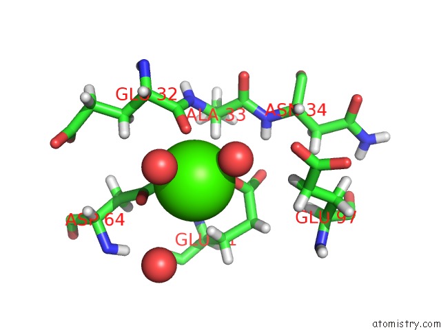
Mono view
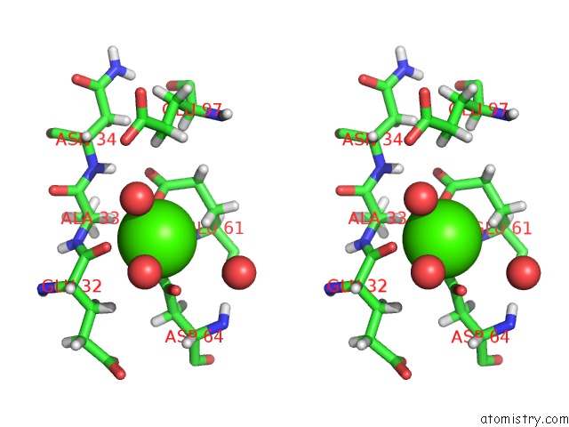
Stereo pair view
Mono view
Stereo pair view
A full contact list of Calcium with other atoms in the Ca binding
site number 1 of Structure of A Hydrolase with An Inhibitor within 5.0Å range:
|
Reference:
N.Cekic,
J.E.Heinonen,
K.A.Stubbs,
C.Roth,
Y.He,
A.J.Bennet,
E.J.Mceachern,
G.J.Davies,
D.J.Vocadlo.
Analysis of Transition State Mimicry By Tight Binding Aminothiazoline Inhibitors Provides Insight Into Catalysis By Humano-Glcnacase. Chem Sci V. 7 3742 2016.
ISSN: ISSN 2041-6520
PubMed: 29997861
DOI: 10.1039/C6SC00370B
Page generated: Sun Jul 14 19:16:56 2024
ISSN: ISSN 2041-6520
PubMed: 29997861
DOI: 10.1039/C6SC00370B
Last articles
Zn in 9J0NZn in 9J0O
Zn in 9J0P
Zn in 9FJX
Zn in 9EKB
Zn in 9C0F
Zn in 9CAH
Zn in 9CH0
Zn in 9CH3
Zn in 9CH1