Calcium »
PDB 5fl0-5g39 »
5fq6 »
Calcium in PDB 5fq6: Crystal Structure of the Suscd Complex BT2261-2264 From Bacteroides Thetaiotaomicron
Protein crystallography data
The structure of Crystal Structure of the Suscd Complex BT2261-2264 From Bacteroides Thetaiotaomicron, PDB code: 5fq6
was solved by
A.J.Glenwright,
K.R.Pothula,
D.S.Chorev,
A.Basle,
C.V.Robinson,
U.Kleinekathoefer,
D.N.Bolam,
B.Van Den Berg,
with X-Ray Crystallography technique. A brief refinement statistics is given in the table below:
| Resolution Low / High (Å) | 48.80 / 2.80 |
| Space group | P 1 21 1 |
| Cell size a, b, c (Å), α, β, γ (°) | 222.027, 91.941, 261.219, 90.00, 98.23, 90.00 |
| R / Rfree (%) | 19.8 / 25.5 |
Other elements in 5fq6:
The structure of Crystal Structure of the Suscd Complex BT2261-2264 From Bacteroides Thetaiotaomicron also contains other interesting chemical elements:
| Sodium | (Na) | 4 atoms |
Calcium Binding Sites:
The binding sites of Calcium atom in the Crystal Structure of the Suscd Complex BT2261-2264 From Bacteroides Thetaiotaomicron
(pdb code 5fq6). This binding sites where shown within
5.0 Angstroms radius around Calcium atom.
In total 4 binding sites of Calcium where determined in the Crystal Structure of the Suscd Complex BT2261-2264 From Bacteroides Thetaiotaomicron, PDB code: 5fq6:
Jump to Calcium binding site number: 1; 2; 3; 4;
In total 4 binding sites of Calcium where determined in the Crystal Structure of the Suscd Complex BT2261-2264 From Bacteroides Thetaiotaomicron, PDB code: 5fq6:
Jump to Calcium binding site number: 1; 2; 3; 4;
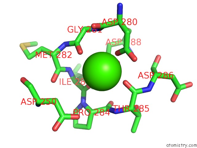
Calcium binding site 1 out of 4 in 5fq6
Go back to
Calcium binding site 1 out
of 4 in the Crystal Structure of the Suscd Complex BT2261-2264 From Bacteroides Thetaiotaomicron
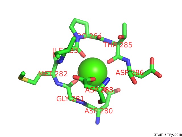
Mono view

Stereo pair view
Mono view
Stereo pair view
A full contact list of Calcium with other atoms in the Ca binding
site number 1 of Crystal Structure of the Suscd Complex BT2261-2264 From Bacteroides Thetaiotaomicron within 5.0Å range:
|
Calcium binding site 2 out of 4 in 5fq6
Go back to
Calcium binding site 2 out
of 4 in the Crystal Structure of the Suscd Complex BT2261-2264 From Bacteroides Thetaiotaomicron
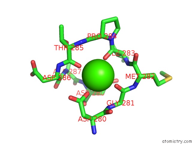
Mono view
Stereo pair view
Mono view
Stereo pair view
A full contact list of Calcium with other atoms in the Ca binding
site number 2 of Crystal Structure of the Suscd Complex BT2261-2264 From Bacteroides Thetaiotaomicron within 5.0Å range:
|
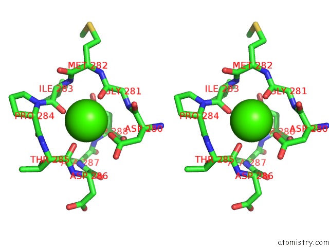
Calcium binding site 3 out of 4 in 5fq6
Go back to
Calcium binding site 3 out
of 4 in the Crystal Structure of the Suscd Complex BT2261-2264 From Bacteroides Thetaiotaomicron

Mono view
Stereo pair view
Mono view
Stereo pair view
A full contact list of Calcium with other atoms in the Ca binding
site number 3 of Crystal Structure of the Suscd Complex BT2261-2264 From Bacteroides Thetaiotaomicron within 5.0Å range:
|
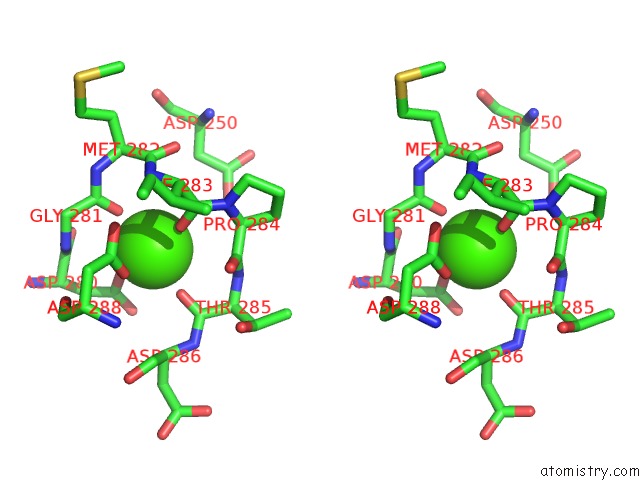
Calcium binding site 4 out of 4 in 5fq6
Go back to
Calcium binding site 4 out
of 4 in the Crystal Structure of the Suscd Complex BT2261-2264 From Bacteroides Thetaiotaomicron
Mono view

Stereo pair view
Mono view
Stereo pair view
A full contact list of Calcium with other atoms in the Ca binding
site number 4 of Crystal Structure of the Suscd Complex BT2261-2264 From Bacteroides Thetaiotaomicron within 5.0Å range:
|
Reference:
A.J.Glenwright,
K.R.Pothula,
S.P.Bhamidimarri,
D.S.Chorev,
A.Basle,
S.J.Firbank,
H.Zheng,
C.V.Robinson,
M.Winterhalter,
U.Kleinekathofer,
D.N.Bolam,
B.Van Den Berg.
Structural Basis For Nutrient Acquisition By Dominant Members of the Human Gut Microbiota. Nature V. 541 407 2017.
ISSN: ESSN 1476-4687
PubMed: 28077872
DOI: 10.1038/NATURE20828
Page generated: Sun Jul 14 19:18:12 2024
ISSN: ESSN 1476-4687
PubMed: 28077872
DOI: 10.1038/NATURE20828
Last articles
Zn in 9J0NZn in 9J0O
Zn in 9J0P
Zn in 9FJX
Zn in 9EKB
Zn in 9C0F
Zn in 9CAH
Zn in 9CH0
Zn in 9CH3
Zn in 9CH1