Calcium »
PDB 5fl0-5g39 »
5fwa »
Calcium in PDB 5fwa: Crystal Structure of Mus Musculus Protein Arginine Methyltransferase 2 with CP1
Enzymatic activity of Crystal Structure of Mus Musculus Protein Arginine Methyltransferase 2 with CP1
All present enzymatic activity of Crystal Structure of Mus Musculus Protein Arginine Methyltransferase 2 with CP1:
2.1.1.125;
2.1.1.125;
Protein crystallography data
The structure of Crystal Structure of Mus Musculus Protein Arginine Methyltransferase 2 with CP1, PDB code: 5fwa
was solved by
V.Cura,
N.Troffer-Charlier,
N.Marechal,
L.Bonnefond,
J.Cavarelli,
with X-Ray Crystallography technique. A brief refinement statistics is given in the table below:
| Resolution Low / High (Å) | 43.37 / 1.80 |
| Space group | C 2 2 21 |
| Cell size a, b, c (Å), α, β, γ (°) | 66.183, 114.676, 132.653, 90.00, 90.00, 90.00 |
| R / Rfree (%) | 17.2 / 19.1 |
Other elements in 5fwa:
The structure of Crystal Structure of Mus Musculus Protein Arginine Methyltransferase 2 with CP1 also contains other interesting chemical elements:
| Chlorine | (Cl) | 1 atom |
Calcium Binding Sites:
The binding sites of Calcium atom in the Crystal Structure of Mus Musculus Protein Arginine Methyltransferase 2 with CP1
(pdb code 5fwa). This binding sites where shown within
5.0 Angstroms radius around Calcium atom.
In total 2 binding sites of Calcium where determined in the Crystal Structure of Mus Musculus Protein Arginine Methyltransferase 2 with CP1, PDB code: 5fwa:
Jump to Calcium binding site number: 1; 2;
In total 2 binding sites of Calcium where determined in the Crystal Structure of Mus Musculus Protein Arginine Methyltransferase 2 with CP1, PDB code: 5fwa:
Jump to Calcium binding site number: 1; 2;
Calcium binding site 1 out of 2 in 5fwa
Go back to
Calcium binding site 1 out
of 2 in the Crystal Structure of Mus Musculus Protein Arginine Methyltransferase 2 with CP1
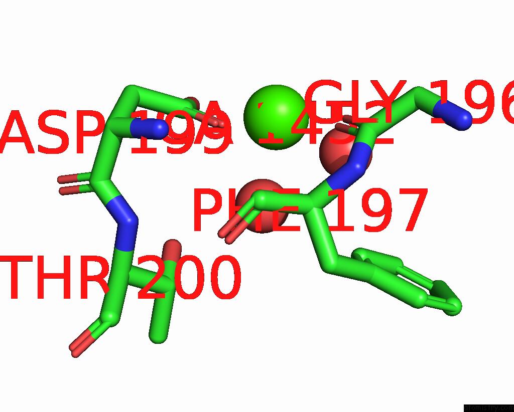
Mono view
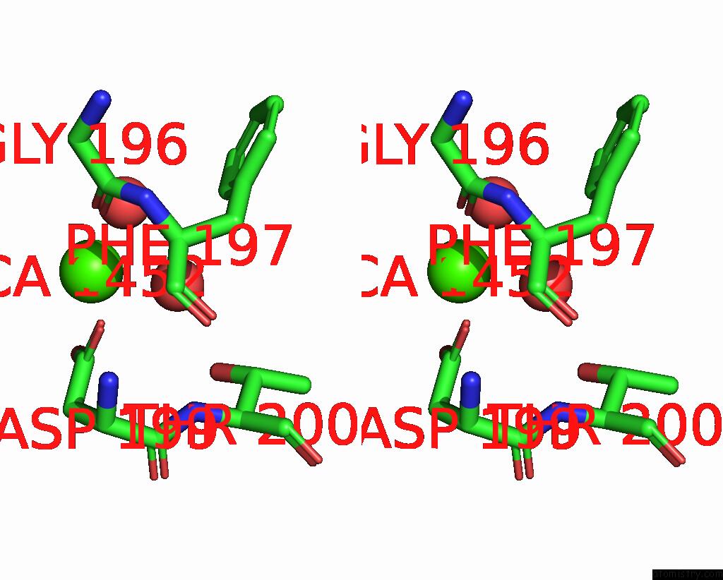
Stereo pair view
Mono view
Stereo pair view
A full contact list of Calcium with other atoms in the Ca binding
site number 1 of Crystal Structure of Mus Musculus Protein Arginine Methyltransferase 2 with CP1 within 5.0Å range:
|
Calcium binding site 2 out of 2 in 5fwa
Go back to
Calcium binding site 2 out
of 2 in the Crystal Structure of Mus Musculus Protein Arginine Methyltransferase 2 with CP1
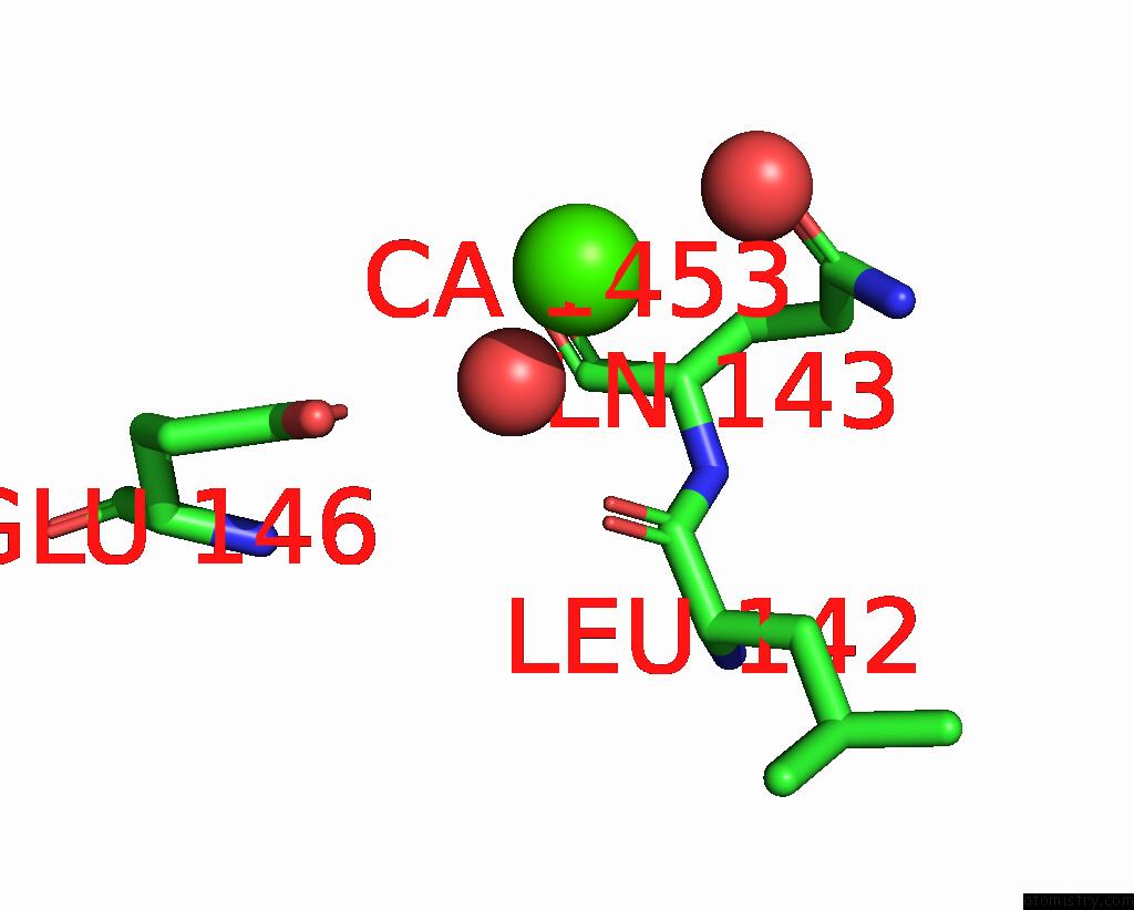
Mono view
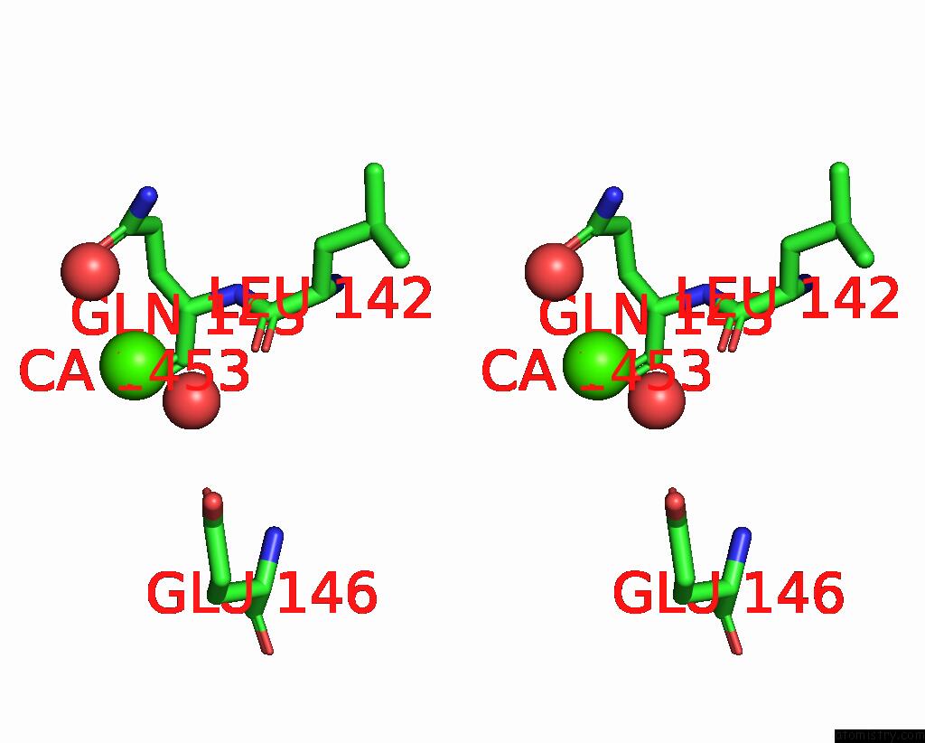
Stereo pair view
Mono view
Stereo pair view
A full contact list of Calcium with other atoms in the Ca binding
site number 2 of Crystal Structure of Mus Musculus Protein Arginine Methyltransferase 2 with CP1 within 5.0Å range:
|
Reference:
V.Cura,
N.Marechal,
N.Troffer-Charlier,
J.M.Strub,
M.J.Van Haren,
N.I.Martin,
S.Cianferani,
L.Bonnefond,
J.Cavarelli.
Structural Studies of Protein Arginine Methyltransferase 2 Reveal Its Interactions with Potential Substrates and Inhibitors. Febs J. V. 284 77 2017.
ISSN: ISSN 1742-4658
PubMed: 27879050
DOI: 10.1111/FEBS.13953
Page generated: Sun Jul 14 19:23:46 2024
ISSN: ISSN 1742-4658
PubMed: 27879050
DOI: 10.1111/FEBS.13953
Last articles
Zn in 9MJ5Zn in 9HNW
Zn in 9G0L
Zn in 9FNE
Zn in 9DZN
Zn in 9E0I
Zn in 9D32
Zn in 9DAK
Zn in 8ZXC
Zn in 8ZUF