Calcium »
PDB 5g3a-5gti »
5gqo »
Calcium in PDB 5gqo: Structure of the Second Single Stranded Dna Binding Protein (Ssbb) From Mycobacterium Smegmatis
Protein crystallography data
The structure of Structure of the Second Single Stranded Dna Binding Protein (Ssbb) From Mycobacterium Smegmatis, PDB code: 5gqo
was solved by
A.Singh,
U.Varshney,
M.Vijayan,
with X-Ray Crystallography technique. A brief refinement statistics is given in the table below:
| Resolution Low / High (Å) | 43.24 / 2.50 |
| Space group | P 65 2 2 |
| Cell size a, b, c (Å), α, β, γ (°) | 73.610, 73.610, 216.210, 90.00, 90.00, 120.00 |
| R / Rfree (%) | 21.1 / 25.6 |
Calcium Binding Sites:
The binding sites of Calcium atom in the Structure of the Second Single Stranded Dna Binding Protein (Ssbb) From Mycobacterium Smegmatis
(pdb code 5gqo). This binding sites where shown within
5.0 Angstroms radius around Calcium atom.
In total only one binding site of Calcium was determined in the Structure of the Second Single Stranded Dna Binding Protein (Ssbb) From Mycobacterium Smegmatis, PDB code: 5gqo:
In total only one binding site of Calcium was determined in the Structure of the Second Single Stranded Dna Binding Protein (Ssbb) From Mycobacterium Smegmatis, PDB code: 5gqo:
Calcium binding site 1 out of 1 in 5gqo
Go back to
Calcium binding site 1 out
of 1 in the Structure of the Second Single Stranded Dna Binding Protein (Ssbb) From Mycobacterium Smegmatis
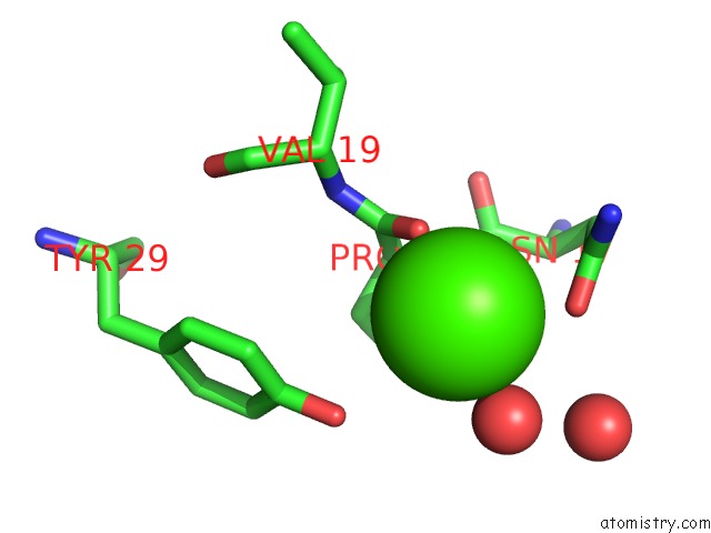
Mono view
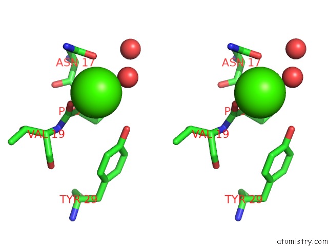
Stereo pair view
Mono view
Stereo pair view
A full contact list of Calcium with other atoms in the Ca binding
site number 1 of Structure of the Second Single Stranded Dna Binding Protein (Ssbb) From Mycobacterium Smegmatis within 5.0Å range:
|
Reference:
A.Singh,
U.Varshney,
M.Vijayan.
Structure of the Second Single Stranded Dna Binding Protein (Ssbb) From Mycobacterium Smegmatis. J. Struct. Biol. V. 196 448 2016.
ISSN: ESSN 1095-8657
PubMed: 27659385
DOI: 10.1016/J.JSB.2016.09.012
Page generated: Sun Jul 14 19:40:51 2024
ISSN: ESSN 1095-8657
PubMed: 27659385
DOI: 10.1016/J.JSB.2016.09.012
Last articles
Zn in 9J0NZn in 9J0O
Zn in 9J0P
Zn in 9FJX
Zn in 9EKB
Zn in 9C0F
Zn in 9CAH
Zn in 9CH0
Zn in 9CH3
Zn in 9CH1