Calcium »
PDB 5gtw-5hca »
5gtw »
Calcium in PDB 5gtw: The N253R Mutant Structures of Trehalose Synthase From Deinococcus Radiodurans Display Two Different Active-Site Conformations
Enzymatic activity of The N253R Mutant Structures of Trehalose Synthase From Deinococcus Radiodurans Display Two Different Active-Site Conformations
All present enzymatic activity of The N253R Mutant Structures of Trehalose Synthase From Deinococcus Radiodurans Display Two Different Active-Site Conformations:
5.4.99.16;
5.4.99.16;
Protein crystallography data
The structure of The N253R Mutant Structures of Trehalose Synthase From Deinococcus Radiodurans Display Two Different Active-Site Conformations, PDB code: 5gtw
was solved by
S.Y.Chow,
Y.J.Wei,
S.H.Liaw,
with X-Ray Crystallography technique. A brief refinement statistics is given in the table below:
| Resolution Low / High (Å) | 20.00 / 2.93 |
| Space group | P 21 21 21 |
| Cell size a, b, c (Å), α, β, γ (°) | 98.383, 134.058, 197.198, 90.00, 90.00, 90.00 |
| R / Rfree (%) | 18.2 / 26.7 |
Other elements in 5gtw:
The structure of The N253R Mutant Structures of Trehalose Synthase From Deinococcus Radiodurans Display Two Different Active-Site Conformations also contains other interesting chemical elements:
| Magnesium | (Mg) | 4 atoms |
Calcium Binding Sites:
The binding sites of Calcium atom in the The N253R Mutant Structures of Trehalose Synthase From Deinococcus Radiodurans Display Two Different Active-Site Conformations
(pdb code 5gtw). This binding sites where shown within
5.0 Angstroms radius around Calcium atom.
In total 4 binding sites of Calcium where determined in the The N253R Mutant Structures of Trehalose Synthase From Deinococcus Radiodurans Display Two Different Active-Site Conformations, PDB code: 5gtw:
Jump to Calcium binding site number: 1; 2; 3; 4;
In total 4 binding sites of Calcium where determined in the The N253R Mutant Structures of Trehalose Synthase From Deinococcus Radiodurans Display Two Different Active-Site Conformations, PDB code: 5gtw:
Jump to Calcium binding site number: 1; 2; 3; 4;
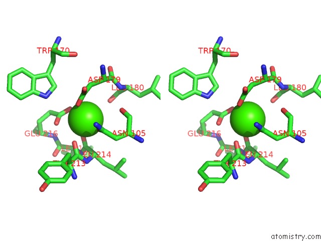
Calcium binding site 1 out of 4 in 5gtw
Go back to
Calcium binding site 1 out
of 4 in the The N253R Mutant Structures of Trehalose Synthase From Deinococcus Radiodurans Display Two Different Active-Site Conformations
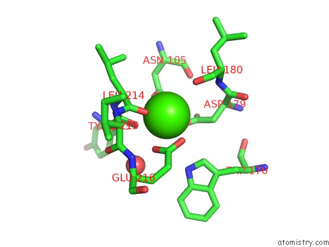
Mono view

Stereo pair view
Mono view
Stereo pair view
A full contact list of Calcium with other atoms in the Ca binding
site number 1 of The N253R Mutant Structures of Trehalose Synthase From Deinococcus Radiodurans Display Two Different Active-Site Conformations within 5.0Å range:
|
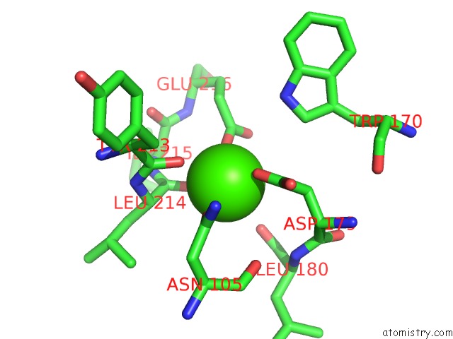
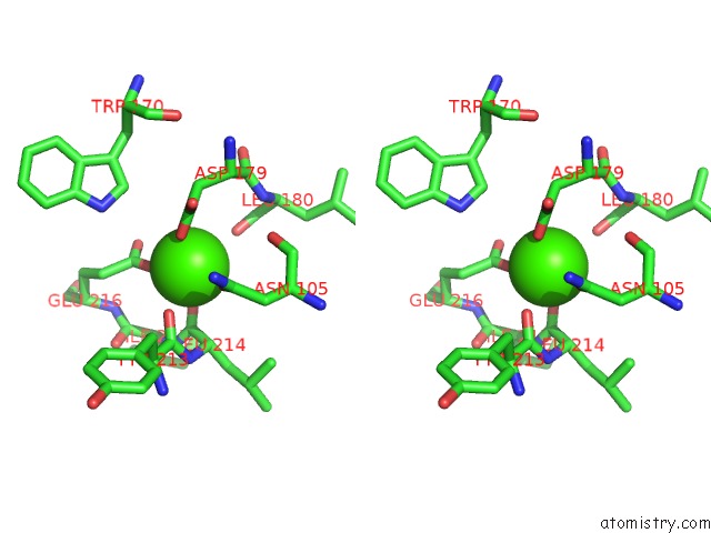
Calcium binding site 2 out of 4 in 5gtw
Go back to
Calcium binding site 2 out
of 4 in the The N253R Mutant Structures of Trehalose Synthase From Deinococcus Radiodurans Display Two Different Active-Site Conformations

Mono view

Stereo pair view
Mono view
Stereo pair view
A full contact list of Calcium with other atoms in the Ca binding
site number 2 of The N253R Mutant Structures of Trehalose Synthase From Deinococcus Radiodurans Display Two Different Active-Site Conformations within 5.0Å range:
|
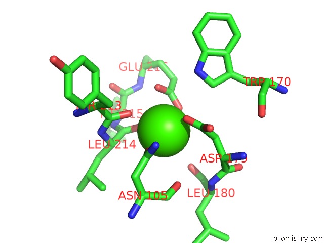
Calcium binding site 3 out of 4 in 5gtw
Go back to
Calcium binding site 3 out
of 4 in the The N253R Mutant Structures of Trehalose Synthase From Deinococcus Radiodurans Display Two Different Active-Site Conformations
Mono view
Stereo pair view
Mono view
Stereo pair view
A full contact list of Calcium with other atoms in the Ca binding
site number 3 of The N253R Mutant Structures of Trehalose Synthase From Deinococcus Radiodurans Display Two Different Active-Site Conformations within 5.0Å range:
|
Calcium binding site 4 out of 4 in 5gtw
Go back to
Calcium binding site 4 out
of 4 in the The N253R Mutant Structures of Trehalose Synthase From Deinococcus Radiodurans Display Two Different Active-Site Conformations
Mono view
Stereo pair view
Mono view
Stereo pair view
A full contact list of Calcium with other atoms in the Ca binding
site number 4 of The N253R Mutant Structures of Trehalose Synthase From Deinococcus Radiodurans Display Two Different Active-Site Conformations within 5.0Å range:
|
Reference:
S.Y.Chow,
Y.J.Wei,
S.H.Liaw.
The N253R Mutant Structures of Trehalose Synthase From Deinococcus Radiodurans Display Two Different Active-Site Conformations To Be Published.
Page generated: Wed Jul 9 06:16:36 2025
Last articles
F in 4FS2F in 4FV0
F in 4FS1
F in 4FRJ
F in 4FRI
F in 4FOG
F in 4FQ4
F in 4FPH
F in 4FNZ
F in 4FOD