Calcium »
PDB 5gtw-5hca »
5h0p »
Calcium in PDB 5h0p: Crystal Structure of Ef-Hand Protein Mutant
Protein crystallography data
The structure of Crystal Structure of Ef-Hand Protein Mutant, PDB code: 5h0p
was solved by
K.R.Park,
J.Y.An,
J.Y.Kang,
J.G.Lee,
H.S.Youn,
Y.Lee,
S.A.Mun,
C.D.Jun,
W.K.Song,
S.H.Eom,
with X-Ray Crystallography technique. A brief refinement statistics is given in the table below:
| Resolution Low / High (Å) | 29.80 / 1.86 |
| Space group | P 21 21 21 |
| Cell size a, b, c (Å), α, β, γ (°) | 36.675, 51.105, 53.650, 90.00, 90.00, 90.00 |
| R / Rfree (%) | 19.6 / 24.6 |
Calcium Binding Sites:
The binding sites of Calcium atom in the Crystal Structure of Ef-Hand Protein Mutant
(pdb code 5h0p). This binding sites where shown within
5.0 Angstroms radius around Calcium atom.
In total 2 binding sites of Calcium where determined in the Crystal Structure of Ef-Hand Protein Mutant, PDB code: 5h0p:
Jump to Calcium binding site number: 1; 2;
In total 2 binding sites of Calcium where determined in the Crystal Structure of Ef-Hand Protein Mutant, PDB code: 5h0p:
Jump to Calcium binding site number: 1; 2;
Calcium binding site 1 out of 2 in 5h0p
Go back to
Calcium binding site 1 out
of 2 in the Crystal Structure of Ef-Hand Protein Mutant
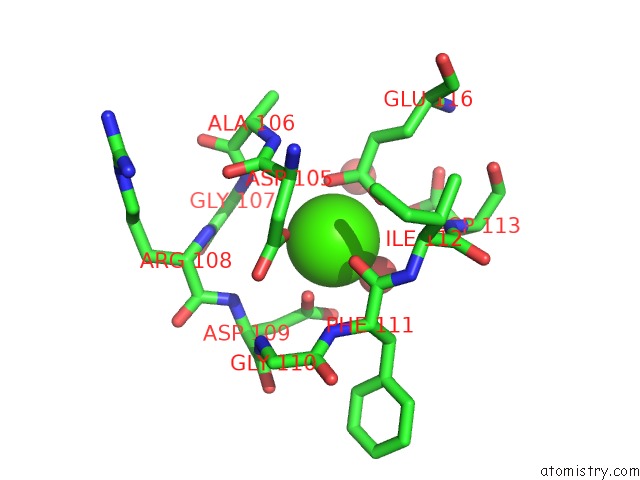
Mono view
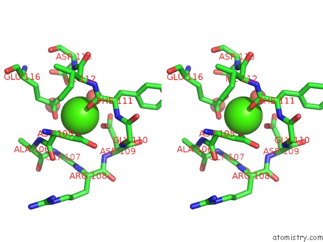
Stereo pair view
Mono view
Stereo pair view
A full contact list of Calcium with other atoms in the Ca binding
site number 1 of Crystal Structure of Ef-Hand Protein Mutant within 5.0Å range:
|
Calcium binding site 2 out of 2 in 5h0p
Go back to
Calcium binding site 2 out
of 2 in the Crystal Structure of Ef-Hand Protein Mutant
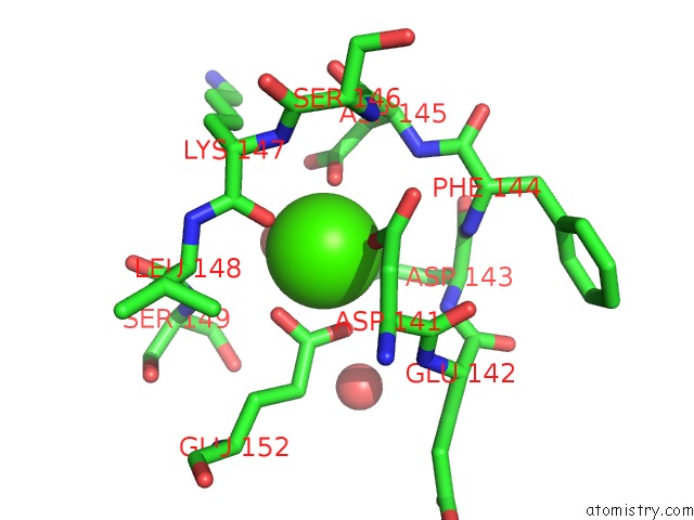
Mono view

Stereo pair view
Mono view
Stereo pair view
A full contact list of Calcium with other atoms in the Ca binding
site number 2 of Crystal Structure of Ef-Hand Protein Mutant within 5.0Å range:
|
Reference:
K.R.Park,
J.Y.An,
J.Y.Kang,
J.G.Lee,
Y.Lee,
S.A.Mun,
C.D.Jun,
W.K.Song,
S.H.Eom.
Structural Mechanism Underlying Regulation of Human EFHD2/Swiprosin-1 Actin-Bundling Activity By SER183 Phosphorylation. Biochem. Biophys. Res. V. 483 442 2017COMMUN..
ISSN: ESSN 1090-2104
PubMed: 28011271
DOI: 10.1016/J.BBRC.2016.12.124
Page generated: Sun Jul 14 19:49:45 2024
ISSN: ESSN 1090-2104
PubMed: 28011271
DOI: 10.1016/J.BBRC.2016.12.124
Last articles
Zn in 9MJ5Zn in 9HNW
Zn in 9G0L
Zn in 9FNE
Zn in 9DZN
Zn in 9E0I
Zn in 9D32
Zn in 9DAK
Zn in 8ZXC
Zn in 8ZUF