Calcium »
PDB 5gtw-5hca »
5h3z »
Calcium in PDB 5h3z: Crystal Structure of 1,2-Beta-Oligoglucan Phosphorylase From Lachnoclostridium Phytofermentans
Enzymatic activity of Crystal Structure of 1,2-Beta-Oligoglucan Phosphorylase From Lachnoclostridium Phytofermentans
All present enzymatic activity of Crystal Structure of 1,2-Beta-Oligoglucan Phosphorylase From Lachnoclostridium Phytofermentans:
2.4.1.333;
2.4.1.333;
Protein crystallography data
The structure of Crystal Structure of 1,2-Beta-Oligoglucan Phosphorylase From Lachnoclostridium Phytofermentans, PDB code: 5h3z
was solved by
M.Nakajima,
N.Tanaka,
N.Furukawa,
T.Nihira,
Y.Kodutsumi,
Y.Takahashi,
N.Sugimoto,
A.Miyanaga,
S.Fushinobu,
H.Taguchi,
H.Nakai,
with X-Ray Crystallography technique. A brief refinement statistics is given in the table below:
| Resolution Low / High (Å) | 42.72 / 2.00 |
| Space group | P 1 21 1 |
| Cell size a, b, c (Å), α, β, γ (°) | 87.761, 94.784, 157.943, 90.00, 98.41, 90.00 |
| R / Rfree (%) | 16.8 / 20.4 |
Calcium Binding Sites:
The binding sites of Calcium atom in the Crystal Structure of 1,2-Beta-Oligoglucan Phosphorylase From Lachnoclostridium Phytofermentans
(pdb code 5h3z). This binding sites where shown within
5.0 Angstroms radius around Calcium atom.
In total 3 binding sites of Calcium where determined in the Crystal Structure of 1,2-Beta-Oligoglucan Phosphorylase From Lachnoclostridium Phytofermentans, PDB code: 5h3z:
Jump to Calcium binding site number: 1; 2; 3;
In total 3 binding sites of Calcium where determined in the Crystal Structure of 1,2-Beta-Oligoglucan Phosphorylase From Lachnoclostridium Phytofermentans, PDB code: 5h3z:
Jump to Calcium binding site number: 1; 2; 3;
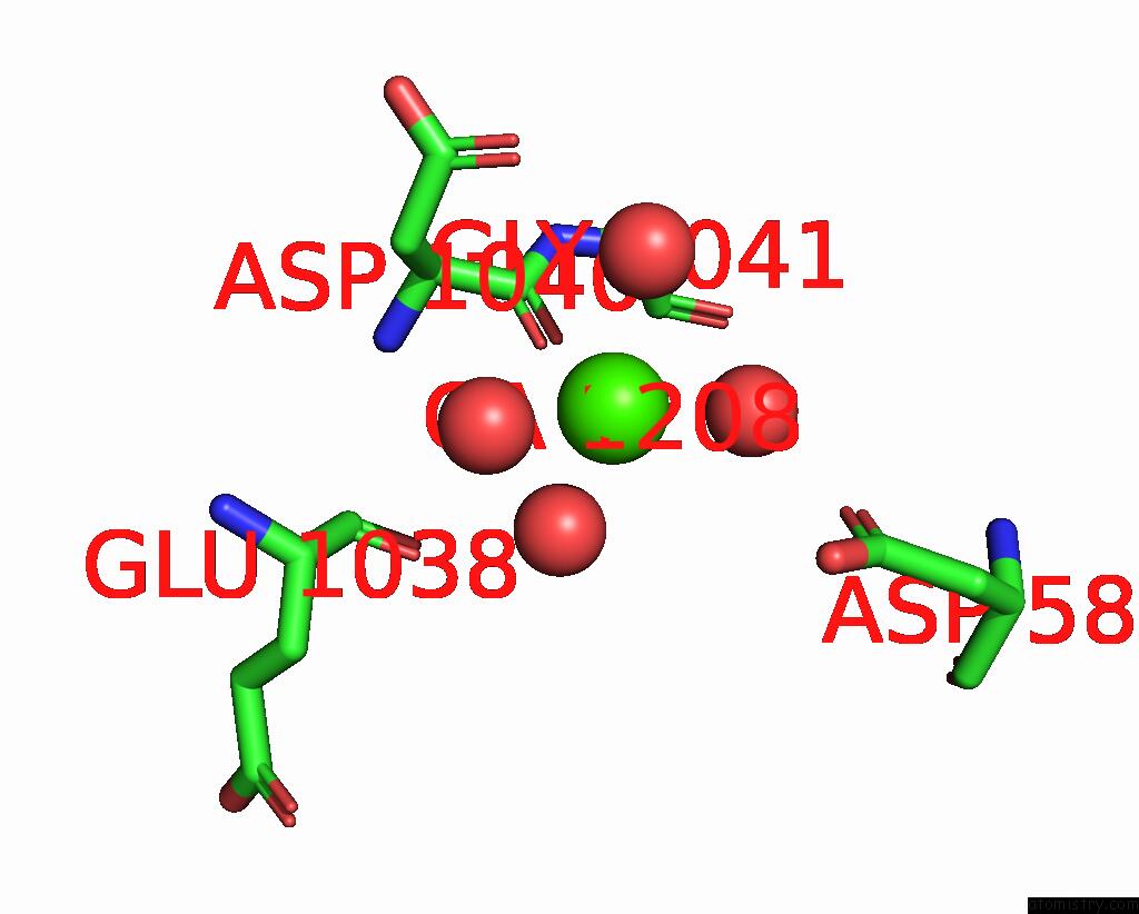
Calcium binding site 1 out of 3 in 5h3z
Go back to
Calcium binding site 1 out
of 3 in the Crystal Structure of 1,2-Beta-Oligoglucan Phosphorylase From Lachnoclostridium Phytofermentans

Mono view

Stereo pair view
Mono view
Stereo pair view
A full contact list of Calcium with other atoms in the Ca binding
site number 1 of Crystal Structure of 1,2-Beta-Oligoglucan Phosphorylase From Lachnoclostridium Phytofermentans within 5.0Å range:
|
Calcium binding site 2 out of 3 in 5h3z
Go back to
Calcium binding site 2 out
of 3 in the Crystal Structure of 1,2-Beta-Oligoglucan Phosphorylase From Lachnoclostridium Phytofermentans

Mono view

Stereo pair view
Mono view
Stereo pair view
A full contact list of Calcium with other atoms in the Ca binding
site number 2 of Crystal Structure of 1,2-Beta-Oligoglucan Phosphorylase From Lachnoclostridium Phytofermentans within 5.0Å range:
|
Calcium binding site 3 out of 3 in 5h3z
Go back to
Calcium binding site 3 out
of 3 in the Crystal Structure of 1,2-Beta-Oligoglucan Phosphorylase From Lachnoclostridium Phytofermentans
Mono view

Stereo pair view
Mono view
Stereo pair view
A full contact list of Calcium with other atoms in the Ca binding
site number 3 of Crystal Structure of 1,2-Beta-Oligoglucan Phosphorylase From Lachnoclostridium Phytofermentans within 5.0Å range:
|
Reference:
M.Nakajima,
N.Tanaka,
N.Furukawa,
T.Nihira,
Y.Kodutsumi,
Y.Takahashi,
N.Sugimoto,
A.Miyanaga,
S.Fushinobu,
H.Taguchi,
H.Nakai.
Mechanistic Insight Into the Substrate Specificity of 1,2-Beta-Oligoglucan Phosphorylase From Lachnoclostridium Phytofermentans Sci Rep V. 7 42671 2017.
ISSN: ESSN 2045-2322
PubMed: 28198470
DOI: 10.1038/SREP42671
Page generated: Sun Jul 14 19:51:09 2024
ISSN: ESSN 2045-2322
PubMed: 28198470
DOI: 10.1038/SREP42671
Last articles
Zn in 9J0NZn in 9J0O
Zn in 9J0P
Zn in 9FJX
Zn in 9EKB
Zn in 9C0F
Zn in 9CAH
Zn in 9CH0
Zn in 9CH3
Zn in 9CH1