Calcium »
PDB 5hcb-5hsa »
5hrt »
Calcium in PDB 5hrt: Crystal Structure of Mouse Autotaxin in Complex with A Dna Aptamer
Enzymatic activity of Crystal Structure of Mouse Autotaxin in Complex with A Dna Aptamer
All present enzymatic activity of Crystal Structure of Mouse Autotaxin in Complex with A Dna Aptamer:
3.1.4.39;
3.1.4.39;
Protein crystallography data
The structure of Crystal Structure of Mouse Autotaxin in Complex with A Dna Aptamer, PDB code: 5hrt
was solved by
K.Kato,
H.Nishimasu,
J.Morita,
R.Ishitani,
O.Nureki,
with X-Ray Crystallography technique. A brief refinement statistics is given in the table below:
| Resolution Low / High (Å) | 45.15 / 2.00 |
| Space group | C 2 2 21 |
| Cell size a, b, c (Å), α, β, γ (°) | 120.062, 208.757, 90.036, 90.00, 90.00, 90.00 |
| R / Rfree (%) | 16.4 / 20.2 |
Other elements in 5hrt:
The structure of Crystal Structure of Mouse Autotaxin in Complex with A Dna Aptamer also contains other interesting chemical elements:
| Potassium | (K) | 1 atom |
| Zinc | (Zn) | 2 atoms |
| Chlorine | (Cl) | 1 atom |
| Sodium | (Na) | 1 atom |
Calcium Binding Sites:
The binding sites of Calcium atom in the Crystal Structure of Mouse Autotaxin in Complex with A Dna Aptamer
(pdb code 5hrt). This binding sites where shown within
5.0 Angstroms radius around Calcium atom.
In total 2 binding sites of Calcium where determined in the Crystal Structure of Mouse Autotaxin in Complex with A Dna Aptamer, PDB code: 5hrt:
Jump to Calcium binding site number: 1; 2;
In total 2 binding sites of Calcium where determined in the Crystal Structure of Mouse Autotaxin in Complex with A Dna Aptamer, PDB code: 5hrt:
Jump to Calcium binding site number: 1; 2;
Calcium binding site 1 out of 2 in 5hrt
Go back to
Calcium binding site 1 out
of 2 in the Crystal Structure of Mouse Autotaxin in Complex with A Dna Aptamer
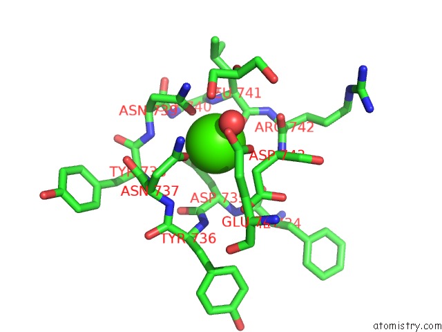
Mono view
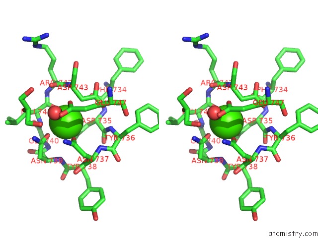
Stereo pair view
Mono view
Stereo pair view
A full contact list of Calcium with other atoms in the Ca binding
site number 1 of Crystal Structure of Mouse Autotaxin in Complex with A Dna Aptamer within 5.0Å range:
|
Calcium binding site 2 out of 2 in 5hrt
Go back to
Calcium binding site 2 out
of 2 in the Crystal Structure of Mouse Autotaxin in Complex with A Dna Aptamer
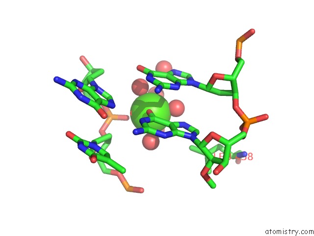
Mono view
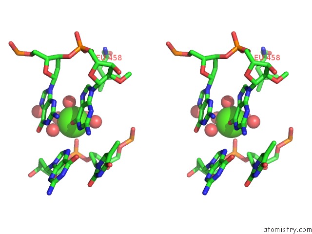
Stereo pair view
Mono view
Stereo pair view
A full contact list of Calcium with other atoms in the Ca binding
site number 2 of Crystal Structure of Mouse Autotaxin in Complex with A Dna Aptamer within 5.0Å range:
|
Reference:
K.Kato,
H.Ikeda,
S.Miyakawa,
S.Futakawa,
Y.Nonaka,
M.Fujiwara,
S.Okudaira,
K.Kano,
J.Aoki,
J.Morita,
R.Ishitani,
H.Nishimasu,
Y.Nakamura,
O.Nureki.
Structural Basis For Specific Inhibition of Autotaxin By A Dna Aptamer Nat.Struct.Mol.Biol. V. 23 395 2016.
ISSN: ESSN 1545-9985
PubMed: 27043297
DOI: 10.1038/NSMB.3200
Page generated: Wed Jul 9 06:34:47 2025
ISSN: ESSN 1545-9985
PubMed: 27043297
DOI: 10.1038/NSMB.3200
Last articles
Cl in 5ICWCl in 5IDY
Cl in 5IEQ
Cl in 5IDN
Cl in 5ICR
Cl in 5IBP
Cl in 5ICU
Cl in 5ICP
Cl in 5IA3
Cl in 5IAE