Calcium »
PDB 5hsq-5i6y »
5i2q »
Calcium in PDB 5i2q: Structure of Ef-Hand Containing Protein
Protein crystallography data
The structure of Structure of Ef-Hand Containing Protein, PDB code: 5i2q
was solved by
K.R.Park,
M.S.Kwon,
J.Y.An,
J.G.Lee,
H.S.Youn,
Y.Lee,
J.Y.Kang,
T.G.Kim,
J.J.Lim,
J.S.Park,
S.H.Lee,
W.K.Song,
H.Cheong,
C.Jun,
S.H.Eom,
with X-Ray Crystallography technique. A brief refinement statistics is given in the table below:
| Resolution Low / High (Å) | 29.39 / 1.94 |
| Space group | P 21 21 21 |
| Cell size a, b, c (Å), α, β, γ (°) | 35.598, 52.107, 55.334, 90.00, 90.00, 90.00 |
| R / Rfree (%) | 17.2 / 20.4 |
Calcium Binding Sites:
The binding sites of Calcium atom in the Structure of Ef-Hand Containing Protein
(pdb code 5i2q). This binding sites where shown within
5.0 Angstroms radius around Calcium atom.
In total only one binding site of Calcium was determined in the Structure of Ef-Hand Containing Protein, PDB code: 5i2q:
In total only one binding site of Calcium was determined in the Structure of Ef-Hand Containing Protein, PDB code: 5i2q:
Calcium binding site 1 out of 1 in 5i2q
Go back to
Calcium binding site 1 out
of 1 in the Structure of Ef-Hand Containing Protein
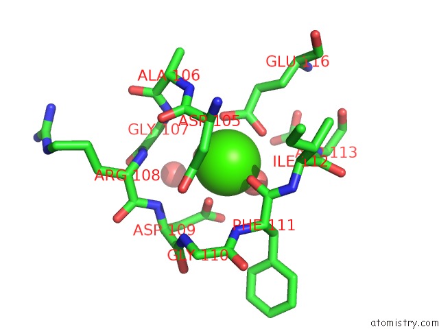
Mono view
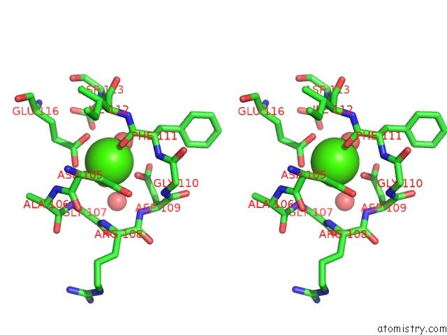
Stereo pair view
Mono view
Stereo pair view
A full contact list of Calcium with other atoms in the Ca binding
site number 1 of Structure of Ef-Hand Containing Protein within 5.0Å range:
|
Reference:
K.R.Park,
M.S.Kwon,
J.Y.An,
J.G.Lee,
H.S.Youn,
Y.Lee,
J.Y.Kang,
T.G.Kim,
J.J.Lim,
J.S.Park,
S.H.Lee,
W.K.Song,
H.K.Cheong,
C.D.Jun,
S.H.Eom.
Structural Implications of Ca(2+)-Dependent Actin-Bundling Function of Human EFHD2/Swiprosin-1. Sci Rep V. 6 39095 2016.
ISSN: ESSN 2045-2322
PubMed: 27974828
DOI: 10.1038/SREP39095
Page generated: Sun Jul 14 20:19:30 2024
ISSN: ESSN 2045-2322
PubMed: 27974828
DOI: 10.1038/SREP39095
Last articles
Zn in 9MJ5Zn in 9HNW
Zn in 9G0L
Zn in 9FNE
Zn in 9DZN
Zn in 9E0I
Zn in 9D32
Zn in 9DAK
Zn in 8ZXC
Zn in 8ZUF