Calcium »
PDB 5i77-5ik8 »
5igb »
Calcium in PDB 5igb: Crystal Structure of Staphylococcal Nuclease Variant Delta+Phs L36D/V66H at Cryogenic Temperature
Enzymatic activity of Crystal Structure of Staphylococcal Nuclease Variant Delta+Phs L36D/V66H at Cryogenic Temperature
All present enzymatic activity of Crystal Structure of Staphylococcal Nuclease Variant Delta+Phs L36D/V66H at Cryogenic Temperature:
3.1.31.1;
3.1.31.1;
Protein crystallography data
The structure of Crystal Structure of Staphylococcal Nuclease Variant Delta+Phs L36D/V66H at Cryogenic Temperature, PDB code: 5igb
was solved by
A.C.Robinson,
A.Theodoru,
J.L.Schlessman,
B.Garcia-Moreno E.,
with X-Ray Crystallography technique. A brief refinement statistics is given in the table below:
| Resolution Low / High (Å) | 38.40 / 1.80 |
| Space group | P 1 21 1 |
| Cell size a, b, c (Å), α, β, γ (°) | 31.139, 60.504, 38.480, 90.00, 93.59, 90.00 |
| R / Rfree (%) | 18.5 / 22.6 |
Calcium Binding Sites:
The binding sites of Calcium atom in the Crystal Structure of Staphylococcal Nuclease Variant Delta+Phs L36D/V66H at Cryogenic Temperature
(pdb code 5igb). This binding sites where shown within
5.0 Angstroms radius around Calcium atom.
In total only one binding site of Calcium was determined in the Crystal Structure of Staphylococcal Nuclease Variant Delta+Phs L36D/V66H at Cryogenic Temperature, PDB code: 5igb:
In total only one binding site of Calcium was determined in the Crystal Structure of Staphylococcal Nuclease Variant Delta+Phs L36D/V66H at Cryogenic Temperature, PDB code: 5igb:
Calcium binding site 1 out of 1 in 5igb
Go back to
Calcium binding site 1 out
of 1 in the Crystal Structure of Staphylococcal Nuclease Variant Delta+Phs L36D/V66H at Cryogenic Temperature
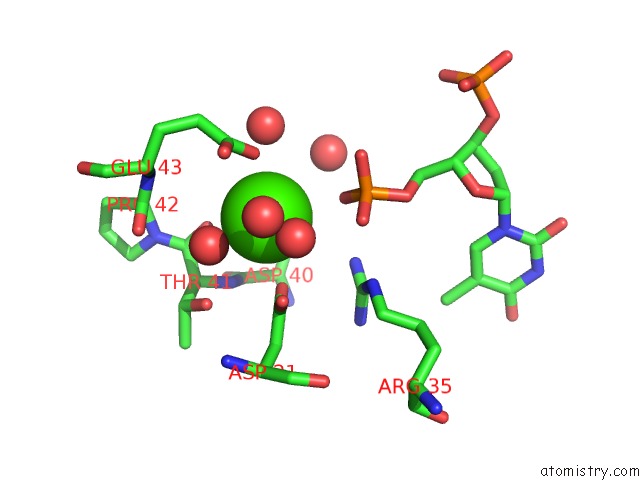
Mono view
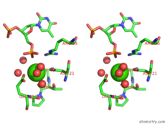
Stereo pair view
Mono view
Stereo pair view
A full contact list of Calcium with other atoms in the Ca binding
site number 1 of Crystal Structure of Staphylococcal Nuclease Variant Delta+Phs L36D/V66H at Cryogenic Temperature within 5.0Å range:
|
Reference:
A.C.Robinson,
A.Theodoru,
J.L.Schlessman,
B.Garcia-Moreno E..
Crystal Structure of Staphylococcal Nuclease Variant Delta+Phs L36D/V66H at Cryogenic Temperature To Be Published.
Page generated: Wed Jul 9 06:47:44 2025
Last articles
Ca in 7GI3Ca in 7G7X
Ca in 7G7V
Ca in 7G7W
Ca in 7G7U
Ca in 7G7T
Ca in 7G7Q
Ca in 7G7S
Ca in 7G7O
Ca in 7G7R