Calcium »
PDB 5i77-5ik8 »
5ijq »
Calcium in PDB 5ijq: Crystal Structure of Autotaxin (ENPP2) Re-Refined
Enzymatic activity of Crystal Structure of Autotaxin (ENPP2) Re-Refined
All present enzymatic activity of Crystal Structure of Autotaxin (ENPP2) Re-Refined:
3.1.4.39;
3.1.4.39;
Protein crystallography data
The structure of Crystal Structure of Autotaxin (ENPP2) Re-Refined, PDB code: 5ijq
was solved by
J.Hausmann,
R.P.Joosten,
A.Perrakis,
with X-Ray Crystallography technique. A brief refinement statistics is given in the table below:
| Resolution Low / High (Å) | 20.00 / 2.05 |
| Space group | P 1 |
| Cell size a, b, c (Å), α, β, γ (°) | 53.808, 63.289, 70.471, 98.78, 106.22, 99.77 |
| R / Rfree (%) | 15.4 / 19.7 |
Other elements in 5ijq:
The structure of Crystal Structure of Autotaxin (ENPP2) Re-Refined also contains other interesting chemical elements:
| Zinc | (Zn) | 2 atoms |
| Iodine | (I) | 25 atoms |
| Sodium | (Na) | 2 atoms |
Calcium Binding Sites:
The binding sites of Calcium atom in the Crystal Structure of Autotaxin (ENPP2) Re-Refined
(pdb code 5ijq). This binding sites where shown within
5.0 Angstroms radius around Calcium atom.
In total only one binding site of Calcium was determined in the Crystal Structure of Autotaxin (ENPP2) Re-Refined, PDB code: 5ijq:
In total only one binding site of Calcium was determined in the Crystal Structure of Autotaxin (ENPP2) Re-Refined, PDB code: 5ijq:
Calcium binding site 1 out of 1 in 5ijq
Go back to
Calcium binding site 1 out
of 1 in the Crystal Structure of Autotaxin (ENPP2) Re-Refined
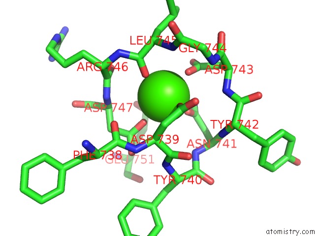
Mono view
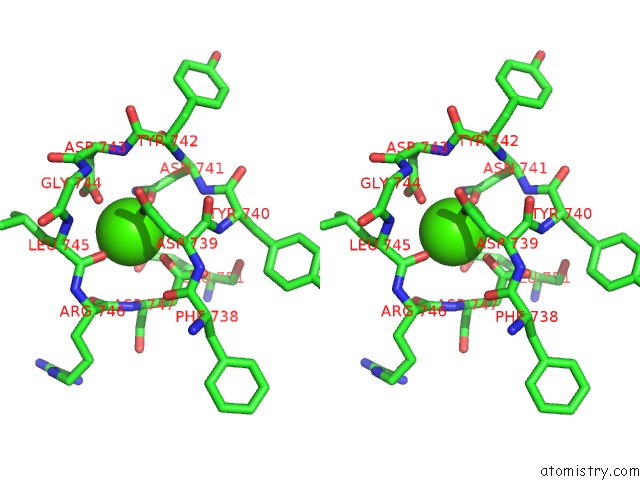
Stereo pair view
Mono view
Stereo pair view
A full contact list of Calcium with other atoms in the Ca binding
site number 1 of Crystal Structure of Autotaxin (ENPP2) Re-Refined within 5.0Å range:
|
Reference:
J.Hausmann,
W.J.Keune,
A.L.Hipgrave Ederveen,
L.Van Zeijl,
R.P.Joosten,
A.Perrakis.
Structural Snapshots of the Catalytic Cycle of the Phosphodiesterase Autotaxin. J.Struct.Biol. V. 195 199 2016.
ISSN: ESSN 1095-8657
PubMed: 27268273
DOI: 10.1016/J.JSB.2016.06.002
Page generated: Sun Jul 14 20:34:01 2024
ISSN: ESSN 1095-8657
PubMed: 27268273
DOI: 10.1016/J.JSB.2016.06.002
Last articles
Zn in 9J0NZn in 9J0O
Zn in 9J0P
Zn in 9FJX
Zn in 9EKB
Zn in 9C0F
Zn in 9CAH
Zn in 9CH0
Zn in 9CH3
Zn in 9CH1