Calcium »
PDB 5iku-5jao »
5iyz »
Calcium in PDB 5iyz: Tubulin-Mmae Complex
Protein crystallography data
The structure of Tubulin-Mmae Complex, PDB code: 5iyz
was solved by
A.B.Waight,
K.Bargsten,
S.Doronina,
M.O.Steinmetz,
D.Sussman,
A.E.Prota,
with X-Ray Crystallography technique. A brief refinement statistics is given in the table below:
| Resolution Low / High (Å) | 71.96 / 1.80 |
| Space group | P 21 21 21 |
| Cell size a, b, c (Å), α, β, γ (°) | 104.510, 156.610, 182.420, 90.00, 90.00, 90.00 |
| R / Rfree (%) | 16.7 / 20.2 |
Other elements in 5iyz:
The structure of Tubulin-Mmae Complex also contains other interesting chemical elements:
| Magnesium | (Mg) | 5 atoms |
Calcium Binding Sites:
The binding sites of Calcium atom in the Tubulin-Mmae Complex
(pdb code 5iyz). This binding sites where shown within
5.0 Angstroms radius around Calcium atom.
In total 2 binding sites of Calcium where determined in the Tubulin-Mmae Complex, PDB code: 5iyz:
Jump to Calcium binding site number: 1; 2;
In total 2 binding sites of Calcium where determined in the Tubulin-Mmae Complex, PDB code: 5iyz:
Jump to Calcium binding site number: 1; 2;
Calcium binding site 1 out of 2 in 5iyz
Go back to
Calcium binding site 1 out
of 2 in the Tubulin-Mmae Complex
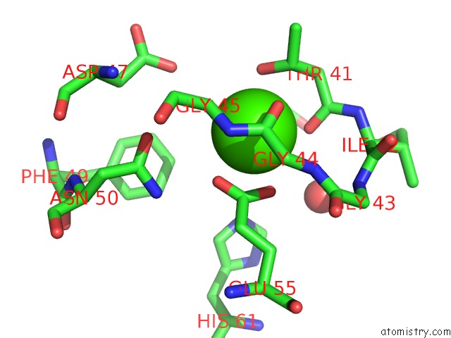
Mono view
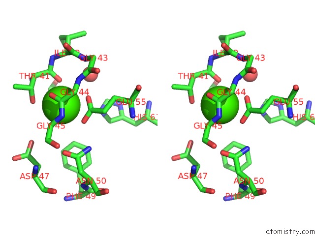
Stereo pair view
Mono view
Stereo pair view
A full contact list of Calcium with other atoms in the Ca binding
site number 1 of Tubulin-Mmae Complex within 5.0Å range:
|
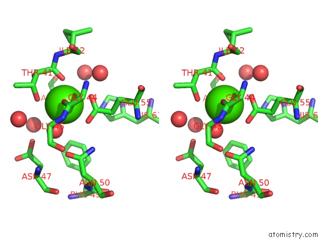
Calcium binding site 2 out of 2 in 5iyz
Go back to
Calcium binding site 2 out
of 2 in the Tubulin-Mmae Complex
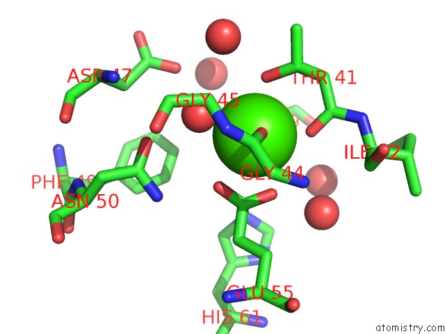
Mono view
Stereo pair view
Mono view
Stereo pair view
A full contact list of Calcium with other atoms in the Ca binding
site number 2 of Tubulin-Mmae Complex within 5.0Å range:
|
Reference:
A.B.Waight,
K.Bargsten,
S.Doronina,
M.O.Steinmetz,
D.Sussman,
A.E.Prota.
Structural Basis of Microtubule Destabilization By Potent Auristatin Anti-Mitotics. Plos One V. 11 60890 2016.
ISSN: ESSN 1932-6203
PubMed: 27518442
DOI: 10.1371/JOURNAL.PONE.0160890
Page generated: Sun Jul 14 20:42:45 2024
ISSN: ESSN 1932-6203
PubMed: 27518442
DOI: 10.1371/JOURNAL.PONE.0160890
Last articles
Zn in 9J0NZn in 9J0O
Zn in 9J0P
Zn in 9FJX
Zn in 9EKB
Zn in 9C0F
Zn in 9CAH
Zn in 9CH0
Zn in 9CH3
Zn in 9CH1