Calcium »
PDB 5iku-5jao »
5j60 »
Calcium in PDB 5j60: Structure of A Thioredoxin Reductase From Gloeobacter Violaceus
Protein crystallography data
The structure of Structure of A Thioredoxin Reductase From Gloeobacter Violaceus, PDB code: 5j60
was solved by
R.M.Buey,
J.M.De Pereda,
M.Balsera,
with X-Ray Crystallography technique. A brief refinement statistics is given in the table below:
| Resolution Low / High (Å) | 49.22 / 1.90 |
| Space group | P 21 21 21 |
| Cell size a, b, c (Å), α, β, γ (°) | 83.278, 122.042, 139.153, 90.00, 90.00, 90.00 |
| R / Rfree (%) | 17.7 / 20.2 |
Calcium Binding Sites:
The binding sites of Calcium atom in the Structure of A Thioredoxin Reductase From Gloeobacter Violaceus
(pdb code 5j60). This binding sites where shown within
5.0 Angstroms radius around Calcium atom.
In total 2 binding sites of Calcium where determined in the Structure of A Thioredoxin Reductase From Gloeobacter Violaceus, PDB code: 5j60:
Jump to Calcium binding site number: 1; 2;
In total 2 binding sites of Calcium where determined in the Structure of A Thioredoxin Reductase From Gloeobacter Violaceus, PDB code: 5j60:
Jump to Calcium binding site number: 1; 2;
Calcium binding site 1 out of 2 in 5j60
Go back to
Calcium binding site 1 out
of 2 in the Structure of A Thioredoxin Reductase From Gloeobacter Violaceus
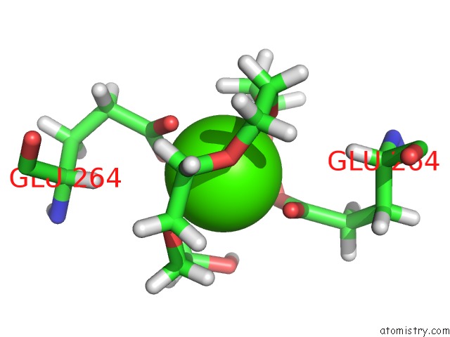
Mono view
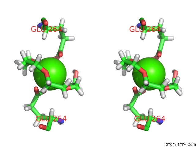
Stereo pair view
Mono view
Stereo pair view
A full contact list of Calcium with other atoms in the Ca binding
site number 1 of Structure of A Thioredoxin Reductase From Gloeobacter Violaceus within 5.0Å range:
|
Calcium binding site 2 out of 2 in 5j60
Go back to
Calcium binding site 2 out
of 2 in the Structure of A Thioredoxin Reductase From Gloeobacter Violaceus
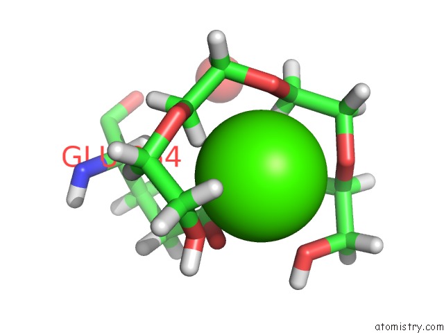
Mono view
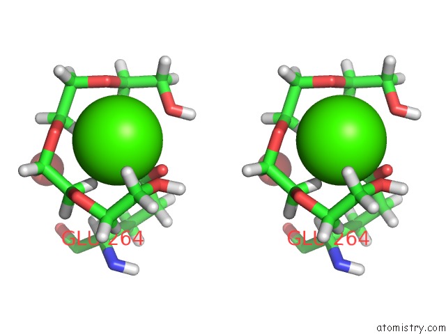
Stereo pair view
Mono view
Stereo pair view
A full contact list of Calcium with other atoms in the Ca binding
site number 2 of Structure of A Thioredoxin Reductase From Gloeobacter Violaceus within 5.0Å range:
|
Reference:
R.M.Buey,
S.Galindo-Trigo,
L.Lopez-Maury,
A.Velazquez-Campoy,
J.L.Revuelta,
F.J.Florencio,
J.M.De Pereda,
P.Schurmann,
B.B.Buchanan,
M.Balsera.
A New Member of the Thioredoxin Reductase Family From Early Oxygenic Photosynthetic Organisms. Mol Plant V. 10 212 2017.
ISSN: ESSN 1752-9867
PubMed: 27418374
DOI: 10.1016/J.MOLP.2016.06.019
Page generated: Wed Jul 9 07:02:17 2025
ISSN: ESSN 1752-9867
PubMed: 27418374
DOI: 10.1016/J.MOLP.2016.06.019
Last articles
Cl in 5KTMCl in 5KSZ
Cl in 5KT1
Cl in 5KSR
Cl in 5KSW
Cl in 5KSS
Cl in 5KSN
Cl in 5KSP
Cl in 5KSK
Cl in 5KRT