Calcium »
PDB 5jap-5jrc »
5jb9 »
Calcium in PDB 5jb9: Crystal Structure of Factor Ixa K98T Variant in Complex with Ppack
Enzymatic activity of Crystal Structure of Factor Ixa K98T Variant in Complex with Ppack
All present enzymatic activity of Crystal Structure of Factor Ixa K98T Variant in Complex with Ppack:
3.4.21.22;
3.4.21.22;
Protein crystallography data
The structure of Crystal Structure of Factor Ixa K98T Variant in Complex with Ppack, PDB code: 5jb9
was solved by
L.H.Kristensen,
H.Brandstetter,
with X-Ray Crystallography technique. A brief refinement statistics is given in the table below:
| Resolution Low / High (Å) | 14.92 / 1.30 |
| Space group | P 21 21 21 |
| Cell size a, b, c (Å), α, β, γ (°) | 44.270, 67.050, 97.150, 90.00, 90.00, 90.00 |
| R / Rfree (%) | 15.1 / 17.6 |
Calcium Binding Sites:
The binding sites of Calcium atom in the Crystal Structure of Factor Ixa K98T Variant in Complex with Ppack
(pdb code 5jb9). This binding sites where shown within
5.0 Angstroms radius around Calcium atom.
In total only one binding site of Calcium was determined in the Crystal Structure of Factor Ixa K98T Variant in Complex with Ppack, PDB code: 5jb9:
In total only one binding site of Calcium was determined in the Crystal Structure of Factor Ixa K98T Variant in Complex with Ppack, PDB code: 5jb9:
Calcium binding site 1 out of 1 in 5jb9
Go back to
Calcium binding site 1 out
of 1 in the Crystal Structure of Factor Ixa K98T Variant in Complex with Ppack
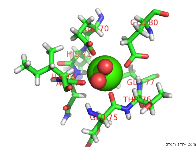
Mono view
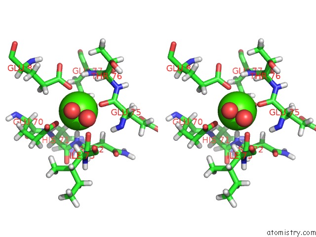
Stereo pair view
Mono view
Stereo pair view
A full contact list of Calcium with other atoms in the Ca binding
site number 1 of Crystal Structure of Factor Ixa K98T Variant in Complex with Ppack within 5.0Å range:
|
Reference:
L.H.Kristensen,
O.H.Olsen,
G.E.Blouse,
H.Brandstetter.
Releasing the Brakes in Coagulation Factor Ixa By Co-Operative Maturation of the Substrate-Binding Site. Biochem.J. V. 473 2395 2016.
ISSN: ESSN 1470-8728
PubMed: 27208168
DOI: 10.1042/BCJ20160336
Page generated: Mon Jul 15 06:25:05 2024
ISSN: ESSN 1470-8728
PubMed: 27208168
DOI: 10.1042/BCJ20160336
Last articles
Zn in 9J0NZn in 9J0O
Zn in 9J0P
Zn in 9FJX
Zn in 9EKB
Zn in 9C0F
Zn in 9CAH
Zn in 9CH0
Zn in 9CH3
Zn in 9CH1