Calcium »
PDB 5jap-5jrc »
5jmo »
Calcium in PDB 5jmo: X-Ray Structure of Furin in Complex with the Inhibitory Antibody NB14
Enzymatic activity of X-Ray Structure of Furin in Complex with the Inhibitory Antibody NB14
All present enzymatic activity of X-Ray Structure of Furin in Complex with the Inhibitory Antibody NB14:
3.4.21.75;
3.4.21.75;
Protein crystallography data
The structure of X-Ray Structure of Furin in Complex with the Inhibitory Antibody NB14, PDB code: 5jmo
was solved by
S.O.Dahms,
M.E.Than,
with X-Ray Crystallography technique. A brief refinement statistics is given in the table below:
| Resolution Low / High (Å) | 66.38 / 2.00 |
| Space group | P 21 2 21 |
| Cell size a, b, c (Å), α, β, γ (°) | 169.775, 50.039, 144.244, 90.00, 90.00, 90.00 |
| R / Rfree (%) | 16.3 / 19.7 |
Other elements in 5jmo:
The structure of X-Ray Structure of Furin in Complex with the Inhibitory Antibody NB14 also contains other interesting chemical elements:
| Sodium | (Na) | 2 atoms |
Calcium Binding Sites:
The binding sites of Calcium atom in the X-Ray Structure of Furin in Complex with the Inhibitory Antibody NB14
(pdb code 5jmo). This binding sites where shown within
5.0 Angstroms radius around Calcium atom.
In total 6 binding sites of Calcium where determined in the X-Ray Structure of Furin in Complex with the Inhibitory Antibody NB14, PDB code: 5jmo:
Jump to Calcium binding site number: 1; 2; 3; 4; 5; 6;
In total 6 binding sites of Calcium where determined in the X-Ray Structure of Furin in Complex with the Inhibitory Antibody NB14, PDB code: 5jmo:
Jump to Calcium binding site number: 1; 2; 3; 4; 5; 6;
Calcium binding site 1 out of 6 in 5jmo
Go back to
Calcium binding site 1 out
of 6 in the X-Ray Structure of Furin in Complex with the Inhibitory Antibody NB14

Mono view
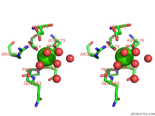
Stereo pair view
Mono view
Stereo pair view
A full contact list of Calcium with other atoms in the Ca binding
site number 1 of X-Ray Structure of Furin in Complex with the Inhibitory Antibody NB14 within 5.0Å range:
|
Calcium binding site 2 out of 6 in 5jmo
Go back to
Calcium binding site 2 out
of 6 in the X-Ray Structure of Furin in Complex with the Inhibitory Antibody NB14

Mono view

Stereo pair view
Mono view
Stereo pair view
A full contact list of Calcium with other atoms in the Ca binding
site number 2 of X-Ray Structure of Furin in Complex with the Inhibitory Antibody NB14 within 5.0Å range:
|
Calcium binding site 3 out of 6 in 5jmo
Go back to
Calcium binding site 3 out
of 6 in the X-Ray Structure of Furin in Complex with the Inhibitory Antibody NB14

Mono view

Stereo pair view
Mono view
Stereo pair view
A full contact list of Calcium with other atoms in the Ca binding
site number 3 of X-Ray Structure of Furin in Complex with the Inhibitory Antibody NB14 within 5.0Å range:
|
Calcium binding site 4 out of 6 in 5jmo
Go back to
Calcium binding site 4 out
of 6 in the X-Ray Structure of Furin in Complex with the Inhibitory Antibody NB14

Mono view

Stereo pair view
Mono view
Stereo pair view
A full contact list of Calcium with other atoms in the Ca binding
site number 4 of X-Ray Structure of Furin in Complex with the Inhibitory Antibody NB14 within 5.0Å range:
|
Calcium binding site 5 out of 6 in 5jmo
Go back to
Calcium binding site 5 out
of 6 in the X-Ray Structure of Furin in Complex with the Inhibitory Antibody NB14

Mono view

Stereo pair view
Mono view
Stereo pair view
A full contact list of Calcium with other atoms in the Ca binding
site number 5 of X-Ray Structure of Furin in Complex with the Inhibitory Antibody NB14 within 5.0Å range:
|
Calcium binding site 6 out of 6 in 5jmo
Go back to
Calcium binding site 6 out
of 6 in the X-Ray Structure of Furin in Complex with the Inhibitory Antibody NB14

Mono view

Stereo pair view
Mono view
Stereo pair view
A full contact list of Calcium with other atoms in the Ca binding
site number 6 of X-Ray Structure of Furin in Complex with the Inhibitory Antibody NB14 within 5.0Å range:
|
Reference:
S.O.Dahms,
J.W.Creemers,
Y.Schaub,
G.P.Bourenkov,
T.Zogg,
H.Brandstetter,
M.E.Than.
The Structure of A Furin-Antibody Complex Explains Non-Competitive Inhibition By Steric Exclusion of Substrate Conformers. Sci Rep V. 6 34303 2016.
ISSN: ESSN 2045-2322
PubMed: 27670069
DOI: 10.1038/SREP34303
Page generated: Mon Jul 15 06:28:13 2024
ISSN: ESSN 2045-2322
PubMed: 27670069
DOI: 10.1038/SREP34303
Last articles
Zn in 9MJ5Zn in 9HNW
Zn in 9G0L
Zn in 9FNE
Zn in 9DZN
Zn in 9E0I
Zn in 9D32
Zn in 9DAK
Zn in 8ZXC
Zn in 8ZUF