Calcium »
PDB 5jao-5jr4 »
5jn2 »
Calcium in PDB 5jn2: Crystal Structure of TGCDPK1 Bound to NVPACU106
Protein crystallography data
The structure of Crystal Structure of TGCDPK1 Bound to NVPACU106, PDB code: 5jn2
was solved by
M.El Bakkouri,
J.R.Walker,
P.Loppnau,
S.Graslund,
C.Bountra,
C.H.Arrowsmith,
A.M.Edwards,
R.Hui,
D.V.Lovato,
Structural Genomicsconsortium (Sgc),
with X-Ray Crystallography technique. A brief refinement statistics is given in the table below:
| Resolution Low / High (Å) | 48.12 / 2.20 |
| Space group | P 1 21 1 |
| Cell size a, b, c (Å), α, β, γ (°) | 48.142, 72.473, 65.101, 90.00, 98.63, 90.00 |
| R / Rfree (%) | 23.3 / 26.9 |
Calcium Binding Sites:
The binding sites of Calcium atom in the Crystal Structure of TGCDPK1 Bound to NVPACU106
(pdb code 5jn2). This binding sites where shown within
5.0 Angstroms radius around Calcium atom.
In total only one binding site of Calcium was determined in the Crystal Structure of TGCDPK1 Bound to NVPACU106, PDB code: 5jn2:
In total only one binding site of Calcium was determined in the Crystal Structure of TGCDPK1 Bound to NVPACU106, PDB code: 5jn2:
Calcium binding site 1 out of 1 in 5jn2
Go back to
Calcium binding site 1 out
of 1 in the Crystal Structure of TGCDPK1 Bound to NVPACU106
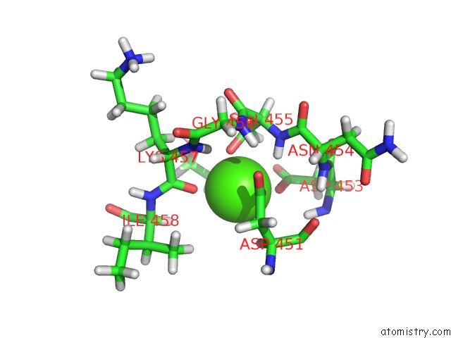
Mono view
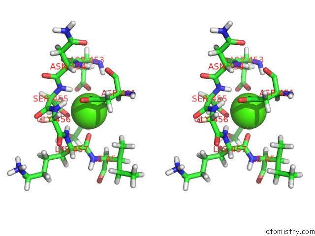
Stereo pair view
Mono view
Stereo pair view
A full contact list of Calcium with other atoms in the Ca binding
site number 1 of Crystal Structure of TGCDPK1 Bound to NVPACU106 within 5.0Å range:
|
Reference:
M.El Bakkouri,
J.R.Walker,
P.Loppnau,
S.Graslund,
C.Bountra,
C.H.Arrowsmith,
A.M.Edwards,
R.Hui,
D.V.Lovato,
Structural Genomics Consortium (Sgc).
Crystal Structure of TGCDPK1 Bound to NVPACU106 To Be Published.
Page generated: Mon Jul 15 06:28:58 2024
Last articles
Zn in 9JYWZn in 9IR4
Zn in 9IR3
Zn in 9GMX
Zn in 9GMW
Zn in 9JEJ
Zn in 9ERF
Zn in 9ERE
Zn in 9EGV
Zn in 9EGW