Calcium »
PDB 5kai-5klg »
5kj8 »
Calcium in PDB 5kj8: Structure of the CA2+-Bound Synaptotagmin-1 Snare Complex (Long Unit Cell Form) - From Synchrotron Diffraction
Protein crystallography data
The structure of Structure of the CA2+-Bound Synaptotagmin-1 Snare Complex (Long Unit Cell Form) - From Synchrotron Diffraction, PDB code: 5kj8
was solved by
A.Y.Lyubimov,
M.Uervirojnangkoorn,
Q.Zhou,
M.Zhao,
N.K.Sauter,
A.S.Brewster,
W.I.Weis,
A.T.Brunger,
with X-Ray Crystallography technique. A brief refinement statistics is given in the table below:
| Resolution Low / High (Å) | 49.64 / 4.10 |
| Space group | P 21 21 21 |
| Cell size a, b, c (Å), α, β, γ (°) | 68.790, 169.710, 286.790, 90.00, 90.00, 90.00 |
| R / Rfree (%) | 28.3 / 29.7 |
Calcium Binding Sites:
Pages:
>>> Page 1 <<< Page 2, Binding sites: 11 - 14;Binding sites:
The binding sites of Calcium atom in the Structure of the CA2+-Bound Synaptotagmin-1 Snare Complex (Long Unit Cell Form) - From Synchrotron Diffraction (pdb code 5kj8). This binding sites where shown within 5.0 Angstroms radius around Calcium atom.In total 14 binding sites of Calcium where determined in the Structure of the CA2+-Bound Synaptotagmin-1 Snare Complex (Long Unit Cell Form) - From Synchrotron Diffraction, PDB code: 5kj8:
Jump to Calcium binding site number: 1; 2; 3; 4; 5; 6; 7; 8; 9; 10;
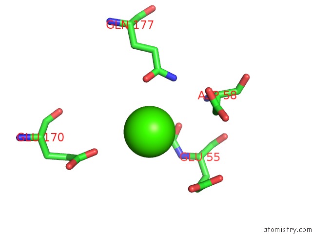
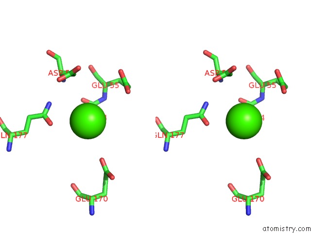
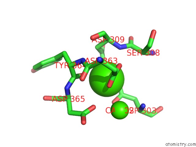
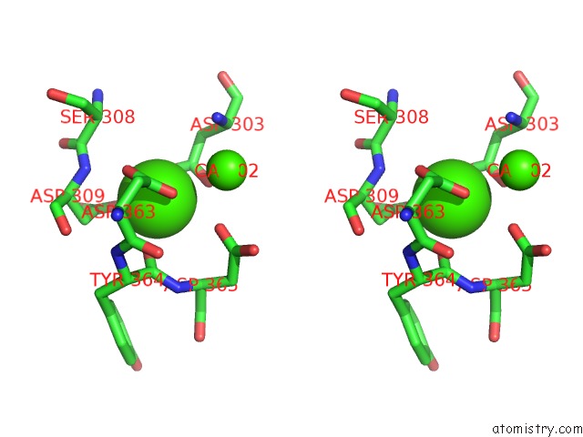
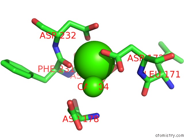
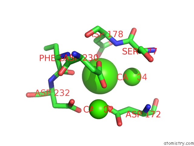
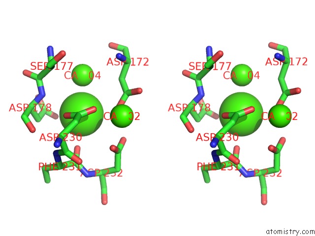
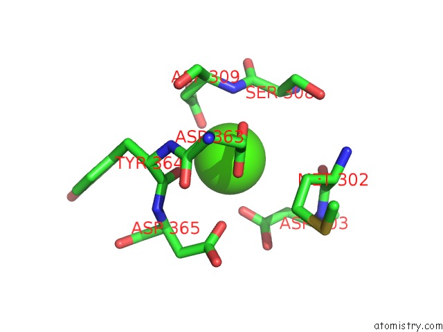
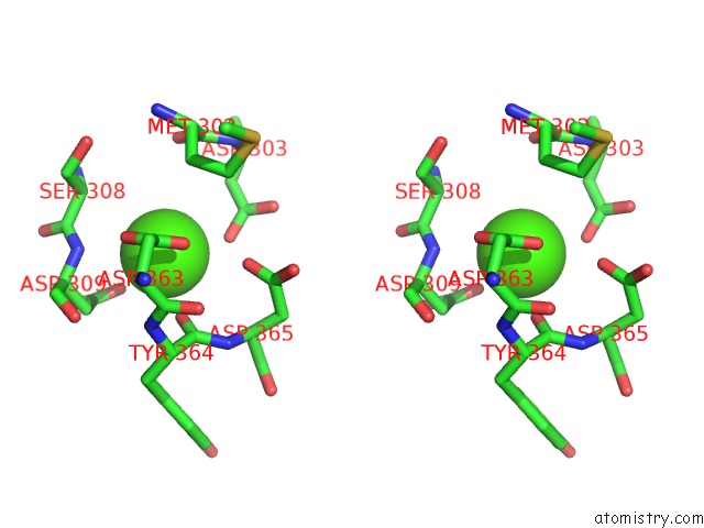
Calcium binding site 1 out of 14 in 5kj8
Go back to
Calcium binding site 1 out
of 14 in the Structure of the CA2+-Bound Synaptotagmin-1 Snare Complex (Long Unit Cell Form) - From Synchrotron Diffraction
Mono view
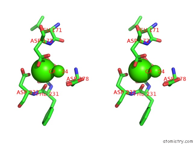
Stereo pair view
Mono view
Stereo pair view
A full contact list of Calcium with other atoms in the Ca binding
site number 1 of Structure of the CA2+-Bound Synaptotagmin-1 Snare Complex (Long Unit Cell Form) - From Synchrotron Diffraction within 5.0Å range:
|
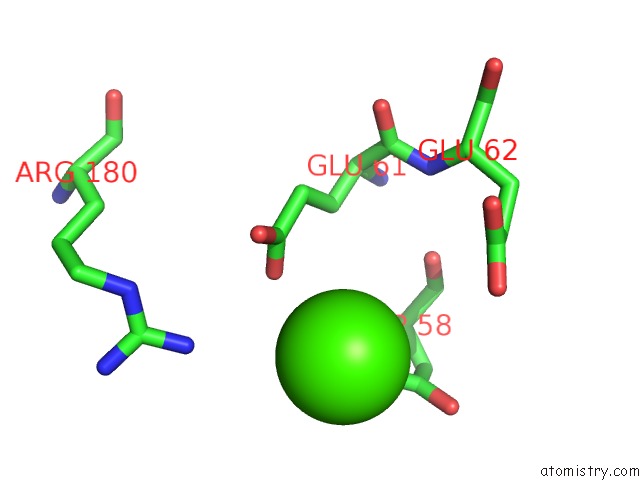
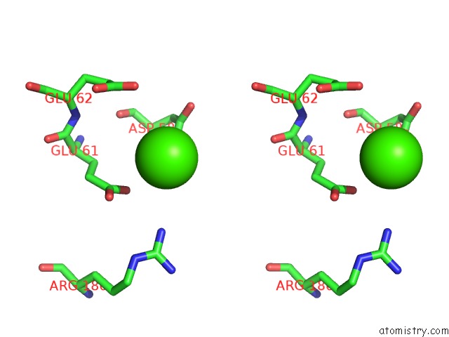
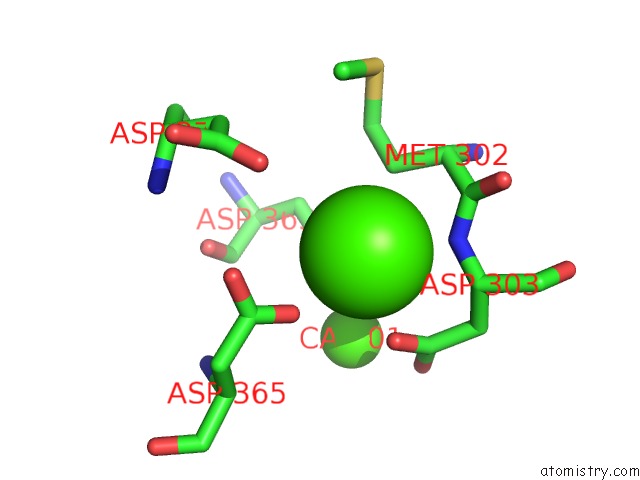
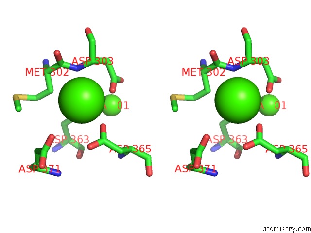
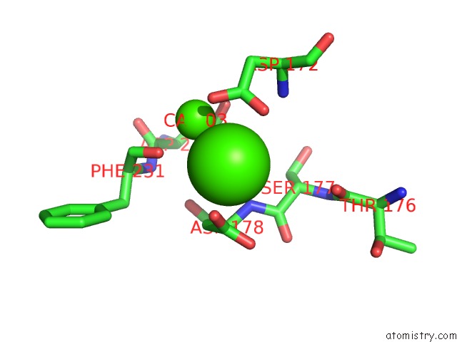
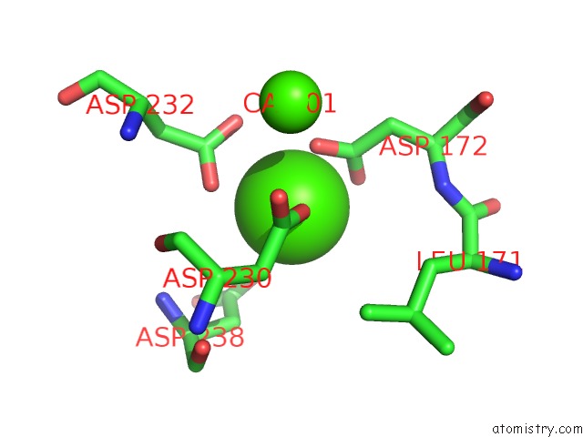
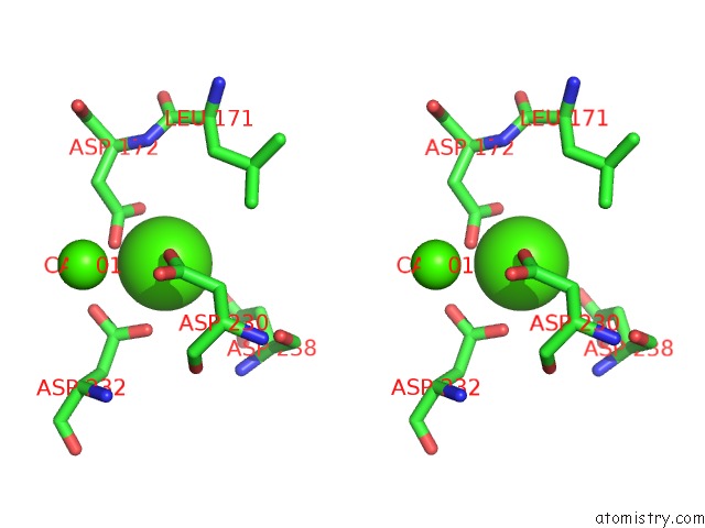
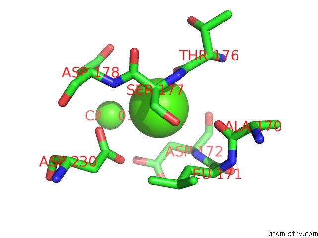
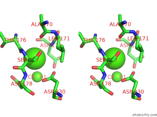
Calcium binding site 2 out of 14 in 5kj8
Go back to
Calcium binding site 2 out
of 14 in the Structure of the CA2+-Bound Synaptotagmin-1 Snare Complex (Long Unit Cell Form) - From Synchrotron Diffraction
Mono view
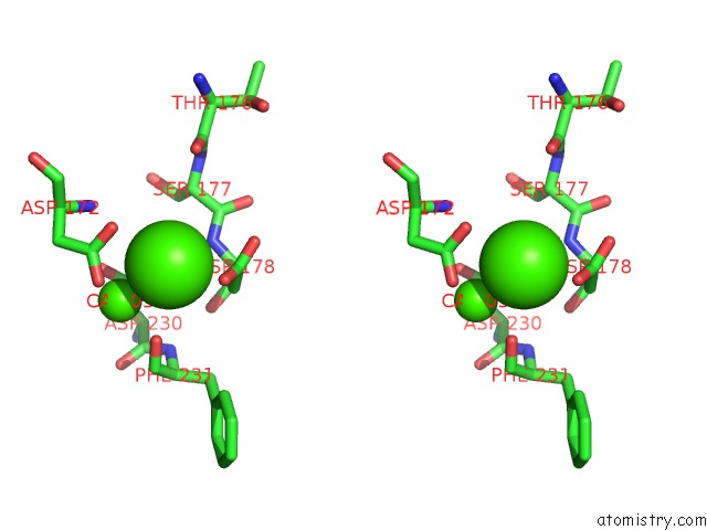
Stereo pair view
Mono view
Stereo pair view
A full contact list of Calcium with other atoms in the Ca binding
site number 2 of Structure of the CA2+-Bound Synaptotagmin-1 Snare Complex (Long Unit Cell Form) - From Synchrotron Diffraction within 5.0Å range:
|
Calcium binding site 3 out of 14 in 5kj8
Go back to
Calcium binding site 3 out
of 14 in the Structure of the CA2+-Bound Synaptotagmin-1 Snare Complex (Long Unit Cell Form) - From Synchrotron Diffraction
Mono view
Stereo pair view
Mono view
Stereo pair view
A full contact list of Calcium with other atoms in the Ca binding
site number 3 of Structure of the CA2+-Bound Synaptotagmin-1 Snare Complex (Long Unit Cell Form) - From Synchrotron Diffraction within 5.0Å range:
|
Calcium binding site 4 out of 14 in 5kj8
Go back to
Calcium binding site 4 out
of 14 in the Structure of the CA2+-Bound Synaptotagmin-1 Snare Complex (Long Unit Cell Form) - From Synchrotron Diffraction
Mono view
Stereo pair view
Mono view
Stereo pair view
A full contact list of Calcium with other atoms in the Ca binding
site number 4 of Structure of the CA2+-Bound Synaptotagmin-1 Snare Complex (Long Unit Cell Form) - From Synchrotron Diffraction within 5.0Å range:
|
Calcium binding site 5 out of 14 in 5kj8
Go back to
Calcium binding site 5 out
of 14 in the Structure of the CA2+-Bound Synaptotagmin-1 Snare Complex (Long Unit Cell Form) - From Synchrotron Diffraction
Mono view
Stereo pair view
Mono view
Stereo pair view
A full contact list of Calcium with other atoms in the Ca binding
site number 5 of Structure of the CA2+-Bound Synaptotagmin-1 Snare Complex (Long Unit Cell Form) - From Synchrotron Diffraction within 5.0Å range:
|
Calcium binding site 6 out of 14 in 5kj8
Go back to
Calcium binding site 6 out
of 14 in the Structure of the CA2+-Bound Synaptotagmin-1 Snare Complex (Long Unit Cell Form) - From Synchrotron Diffraction
Mono view
Stereo pair view
Mono view
Stereo pair view
A full contact list of Calcium with other atoms in the Ca binding
site number 6 of Structure of the CA2+-Bound Synaptotagmin-1 Snare Complex (Long Unit Cell Form) - From Synchrotron Diffraction within 5.0Å range:
|
Calcium binding site 7 out of 14 in 5kj8
Go back to
Calcium binding site 7 out
of 14 in the Structure of the CA2+-Bound Synaptotagmin-1 Snare Complex (Long Unit Cell Form) - From Synchrotron Diffraction
Mono view
Stereo pair view
Mono view
Stereo pair view
A full contact list of Calcium with other atoms in the Ca binding
site number 7 of Structure of the CA2+-Bound Synaptotagmin-1 Snare Complex (Long Unit Cell Form) - From Synchrotron Diffraction within 5.0Å range:
|
Calcium binding site 8 out of 14 in 5kj8
Go back to
Calcium binding site 8 out
of 14 in the Structure of the CA2+-Bound Synaptotagmin-1 Snare Complex (Long Unit Cell Form) - From Synchrotron Diffraction
Mono view
Stereo pair view
Mono view
Stereo pair view
A full contact list of Calcium with other atoms in the Ca binding
site number 8 of Structure of the CA2+-Bound Synaptotagmin-1 Snare Complex (Long Unit Cell Form) - From Synchrotron Diffraction within 5.0Å range:
|
Calcium binding site 9 out of 14 in 5kj8
Go back to
Calcium binding site 9 out
of 14 in the Structure of the CA2+-Bound Synaptotagmin-1 Snare Complex (Long Unit Cell Form) - From Synchrotron Diffraction
Mono view
Stereo pair view
Mono view
Stereo pair view
A full contact list of Calcium with other atoms in the Ca binding
site number 9 of Structure of the CA2+-Bound Synaptotagmin-1 Snare Complex (Long Unit Cell Form) - From Synchrotron Diffraction within 5.0Å range:
|
Calcium binding site 10 out of 14 in 5kj8
Go back to
Calcium binding site 10 out
of 14 in the Structure of the CA2+-Bound Synaptotagmin-1 Snare Complex (Long Unit Cell Form) - From Synchrotron Diffraction
Mono view
Stereo pair view
Mono view
Stereo pair view
A full contact list of Calcium with other atoms in the Ca binding
site number 10 of Structure of the CA2+-Bound Synaptotagmin-1 Snare Complex (Long Unit Cell Form) - From Synchrotron Diffraction within 5.0Å range:
|
Reference:
A.Y.Lyubimov,
M.Uervirojnangkoorn,
O.B.Zeldin,
Q.Zhou,
M.Zhao,
A.S.Brewster,
T.Michels-Clark,
J.M.Holton,
N.K.Sauter,
W.I.Weis,
A.T.Brunger.
Advances in X-Ray Free Electron Laser (Xfel) Diffraction Data Processing Applied to the Crystal Structure of the Synaptotagmin-1 / Snare Complex. Elife V. 5 2016.
ISSN: ESSN 2050-084X
PubMed: 27731796
DOI: 10.7554/ELIFE.18740
Page generated: Mon Jul 15 06:53:25 2024
ISSN: ESSN 2050-084X
PubMed: 27731796
DOI: 10.7554/ELIFE.18740
Last articles
Zn in 9J0NZn in 9J0O
Zn in 9J0P
Zn in 9FJX
Zn in 9EKB
Zn in 9C0F
Zn in 9CAH
Zn in 9CH0
Zn in 9CH3
Zn in 9CH1