Calcium »
PDB 5kai-5klg »
5kk7 »
Calcium in PDB 5kk7: Crystal Structure of the Class I Human Endoplasmic Reticulum 1,2- Alpha-Mannosidase T688A Mutant and Thio-Disaccharide Substrate Analog Complex
Enzymatic activity of Crystal Structure of the Class I Human Endoplasmic Reticulum 1,2- Alpha-Mannosidase T688A Mutant and Thio-Disaccharide Substrate Analog Complex
All present enzymatic activity of Crystal Structure of the Class I Human Endoplasmic Reticulum 1,2- Alpha-Mannosidase T688A Mutant and Thio-Disaccharide Substrate Analog Complex:
3.2.1.113;
3.2.1.113;
Protein crystallography data
The structure of Crystal Structure of the Class I Human Endoplasmic Reticulum 1,2- Alpha-Mannosidase T688A Mutant and Thio-Disaccharide Substrate Analog Complex, PDB code: 5kk7
was solved by
K.Karaveg,
Y.Xiang,
K.W.Moremen,
with X-Ray Crystallography technique. A brief refinement statistics is given in the table below:
| Resolution Low / High (Å) | 42.16 / 1.73 |
| Space group | P 1 |
| Cell size a, b, c (Å), α, β, γ (°) | 53.887, 56.302, 89.555, 105.68, 94.15, 114.25 |
| R / Rfree (%) | 17.6 / 20.9 |
Calcium Binding Sites:
The binding sites of Calcium atom in the Crystal Structure of the Class I Human Endoplasmic Reticulum 1,2- Alpha-Mannosidase T688A Mutant and Thio-Disaccharide Substrate Analog Complex
(pdb code 5kk7). This binding sites where shown within
5.0 Angstroms radius around Calcium atom.
In total 2 binding sites of Calcium where determined in the Crystal Structure of the Class I Human Endoplasmic Reticulum 1,2- Alpha-Mannosidase T688A Mutant and Thio-Disaccharide Substrate Analog Complex, PDB code: 5kk7:
Jump to Calcium binding site number: 1; 2;
In total 2 binding sites of Calcium where determined in the Crystal Structure of the Class I Human Endoplasmic Reticulum 1,2- Alpha-Mannosidase T688A Mutant and Thio-Disaccharide Substrate Analog Complex, PDB code: 5kk7:
Jump to Calcium binding site number: 1; 2;
Calcium binding site 1 out of 2 in 5kk7
Go back to
Calcium binding site 1 out
of 2 in the Crystal Structure of the Class I Human Endoplasmic Reticulum 1,2- Alpha-Mannosidase T688A Mutant and Thio-Disaccharide Substrate Analog Complex
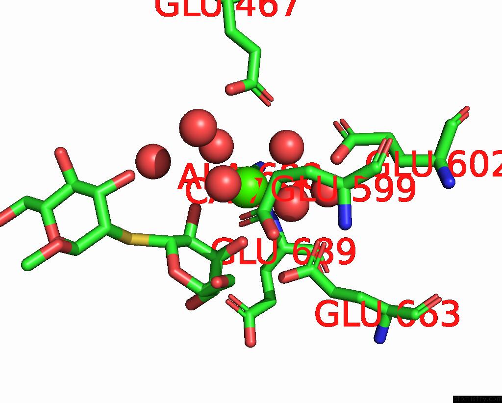
Mono view
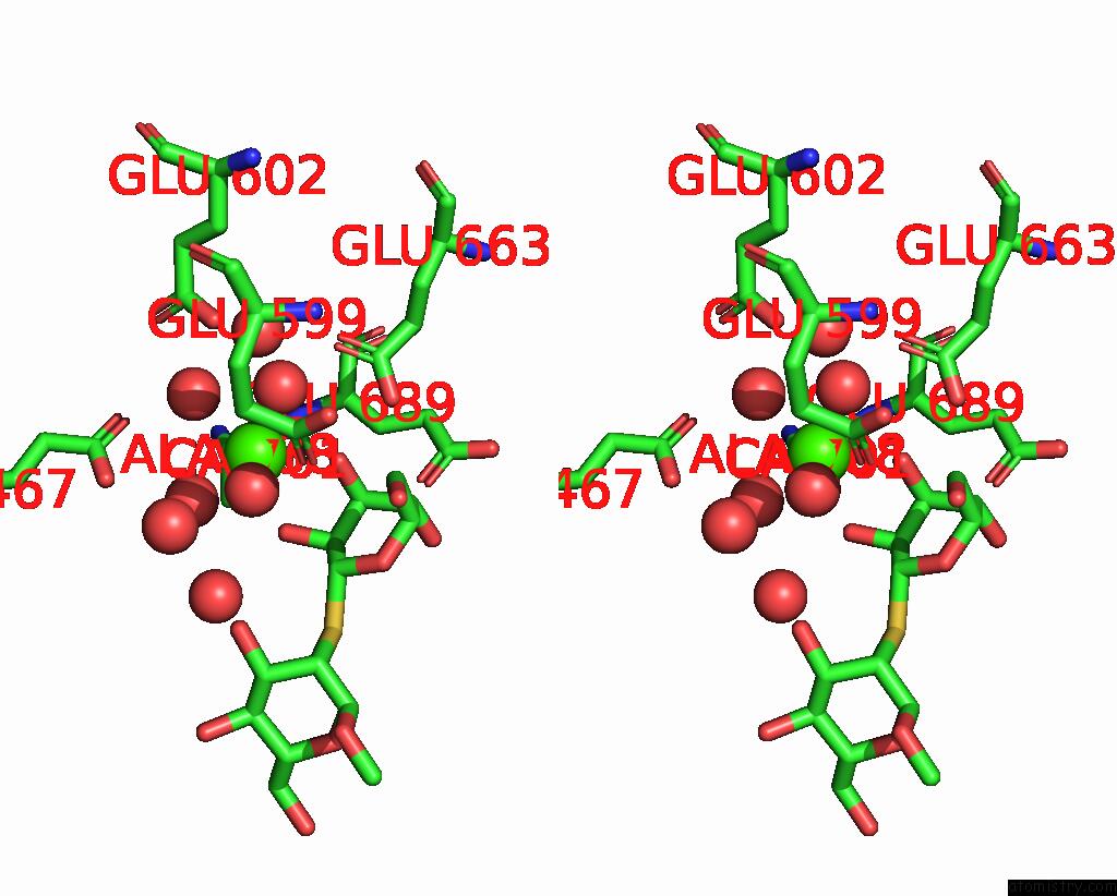
Stereo pair view
Mono view
Stereo pair view
A full contact list of Calcium with other atoms in the Ca binding
site number 1 of Crystal Structure of the Class I Human Endoplasmic Reticulum 1,2- Alpha-Mannosidase T688A Mutant and Thio-Disaccharide Substrate Analog Complex within 5.0Å range:
|
Calcium binding site 2 out of 2 in 5kk7
Go back to
Calcium binding site 2 out
of 2 in the Crystal Structure of the Class I Human Endoplasmic Reticulum 1,2- Alpha-Mannosidase T688A Mutant and Thio-Disaccharide Substrate Analog Complex
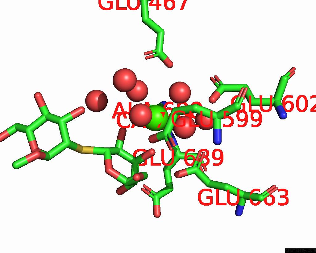
Mono view
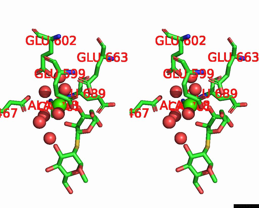
Stereo pair view
Mono view
Stereo pair view
A full contact list of Calcium with other atoms in the Ca binding
site number 2 of Crystal Structure of the Class I Human Endoplasmic Reticulum 1,2- Alpha-Mannosidase T688A Mutant and Thio-Disaccharide Substrate Analog Complex within 5.0Å range:
|
Reference:
Y.Xiang,
K.Karaveg,
K.W.Moremen.
Substrate Recognition and Catalysis By GH47 Alpha-Mannosidases Involved in Asn-Linked Glycan Maturation in the Mammalian Secretory Pathway. Proc. Natl. Acad. Sci. V. 113 E7890 2016U.S.A..
ISSN: ESSN 1091-6490
PubMed: 27856750
DOI: 10.1073/PNAS.1611213113
Page generated: Wed Jul 9 07:26:14 2025
ISSN: ESSN 1091-6490
PubMed: 27856750
DOI: 10.1073/PNAS.1611213113
Last articles
Cl in 5R94Cl in 5R93
Cl in 5R91
Cl in 5R90
Cl in 5R8Z
Cl in 5R8Y
Cl in 5R8X
Cl in 5R8W
Cl in 5R83
Cl in 5R8V