Calcium »
PDB 5kls-5l0u »
5kn8 »
Calcium in PDB 5kn8: Muty N-Terminal Domain in Complex with Undamaged Dna
Protein crystallography data
The structure of Muty N-Terminal Domain in Complex with Undamaged Dna, PDB code: 5kn8
was solved by
L.Wang,
S.Chakravarthy,
G.L.Verdine,
with X-Ray Crystallography technique. A brief refinement statistics is given in the table below:
| Resolution Low / High (Å) | 46.35 / 1.81 |
| Space group | P 1 21 1 |
| Cell size a, b, c (Å), α, β, γ (°) | 49.200, 38.618, 83.796, 90.00, 104.00, 90.00 |
| R / Rfree (%) | 16.7 / 22 |
Other elements in 5kn8:
The structure of Muty N-Terminal Domain in Complex with Undamaged Dna also contains other interesting chemical elements:
| Iron | (Fe) | 4 atoms |
Calcium Binding Sites:
The binding sites of Calcium atom in the Muty N-Terminal Domain in Complex with Undamaged Dna
(pdb code 5kn8). This binding sites where shown within
5.0 Angstroms radius around Calcium atom.
In total only one binding site of Calcium was determined in the Muty N-Terminal Domain in Complex with Undamaged Dna, PDB code: 5kn8:
In total only one binding site of Calcium was determined in the Muty N-Terminal Domain in Complex with Undamaged Dna, PDB code: 5kn8:
Calcium binding site 1 out of 1 in 5kn8
Go back to
Calcium binding site 1 out
of 1 in the Muty N-Terminal Domain in Complex with Undamaged Dna
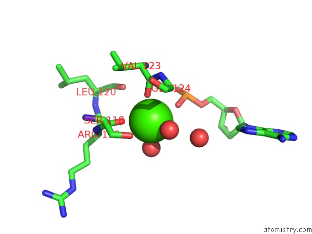
Mono view
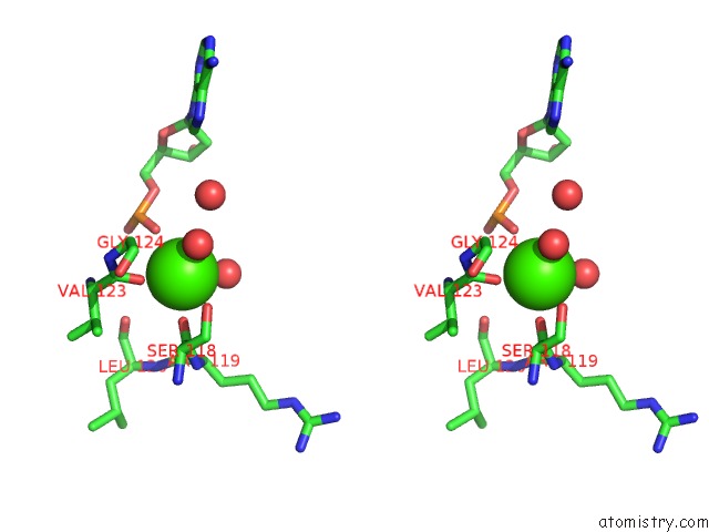
Stereo pair view
Mono view
Stereo pair view
A full contact list of Calcium with other atoms in the Ca binding
site number 1 of Muty N-Terminal Domain in Complex with Undamaged Dna within 5.0Å range:
|
Reference:
L.Wang,
S.Chakravarthy,
G.L.Verdine.
Structural Basis For the Lesion-Scanning Mechanism of the Muty Dna Glycosylase. J. Biol. Chem. V. 292 5007 2017.
ISSN: ESSN 1083-351X
PubMed: 28130451
DOI: 10.1074/JBC.M116.757039
Page generated: Wed Jul 9 07:30:46 2025
ISSN: ESSN 1083-351X
PubMed: 28130451
DOI: 10.1074/JBC.M116.757039
Last articles
Cl in 5R9ACl in 5R99
Cl in 5R98
Cl in 5R96
Cl in 5R97
Cl in 5R95
Cl in 5R92
Cl in 5R94
Cl in 5R93
Cl in 5R91