Calcium »
PDB 5l0v-5lia »
5l0v »
Calcium in PDB 5l0v: Human POGLUT1 in Complex with 2F-Glucose Modified Egf(+) and Udp
Enzymatic activity of Human POGLUT1 in Complex with 2F-Glucose Modified Egf(+) and Udp
All present enzymatic activity of Human POGLUT1 in Complex with 2F-Glucose Modified Egf(+) and Udp:
2.4.2.26;
2.4.2.26;
Protein crystallography data
The structure of Human POGLUT1 in Complex with 2F-Glucose Modified Egf(+) and Udp, PDB code: 5l0v
was solved by
Z.Li,
J.M.Rini,
with X-Ray Crystallography technique. A brief refinement statistics is given in the table below:
| Resolution Low / High (Å) | 35.81 / 1.31 |
| Space group | P 21 21 21 |
| Cell size a, b, c (Å), α, β, γ (°) | 70.810, 74.890, 83.020, 90.00, 90.00, 90.00 |
| R / Rfree (%) | 15.9 / 17.4 |
Other elements in 5l0v:
The structure of Human POGLUT1 in Complex with 2F-Glucose Modified Egf(+) and Udp also contains other interesting chemical elements:
| Fluorine | (F) | 1 atom |
Calcium Binding Sites:
The binding sites of Calcium atom in the Human POGLUT1 in Complex with 2F-Glucose Modified Egf(+) and Udp
(pdb code 5l0v). This binding sites where shown within
5.0 Angstroms radius around Calcium atom.
In total only one binding site of Calcium was determined in the Human POGLUT1 in Complex with 2F-Glucose Modified Egf(+) and Udp, PDB code: 5l0v:
In total only one binding site of Calcium was determined in the Human POGLUT1 in Complex with 2F-Glucose Modified Egf(+) and Udp, PDB code: 5l0v:
Calcium binding site 1 out of 1 in 5l0v
Go back to
Calcium binding site 1 out
of 1 in the Human POGLUT1 in Complex with 2F-Glucose Modified Egf(+) and Udp
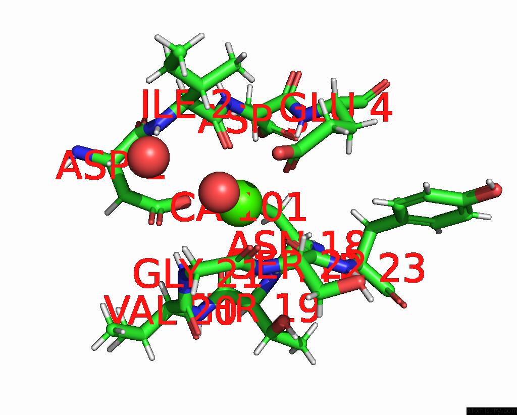
Mono view
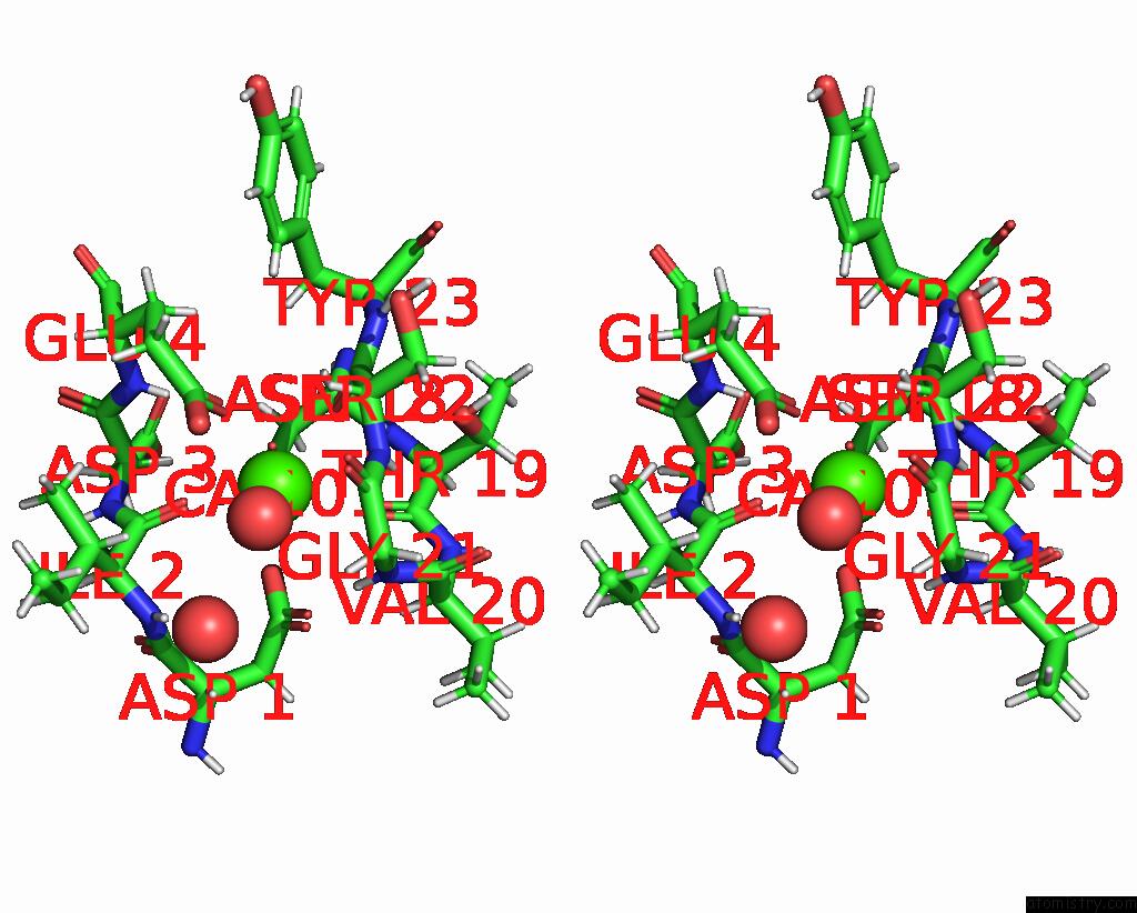
Stereo pair view
Mono view
Stereo pair view
A full contact list of Calcium with other atoms in the Ca binding
site number 1 of Human POGLUT1 in Complex with 2F-Glucose Modified Egf(+) and Udp within 5.0Å range:
|
Reference:
Z.Li,
M.Fischer,
M.Satkunarajah,
D.Zhou,
S.G.Withers,
J.M.Rini.
Structural Basis of Notch O-Glucosylation and O-Xylosylation By Mammalian Protein-O-Glucosyltransferase 1 (POGLUT1). Nat Commun V. 8 185 2017.
ISSN: ESSN 2041-1723
PubMed: 28775322
DOI: 10.1038/S41467-017-00255-7
Page generated: Wed Jul 9 07:41:48 2025
ISSN: ESSN 2041-1723
PubMed: 28775322
DOI: 10.1038/S41467-017-00255-7
Last articles
Fe in 2YXOFe in 2YRS
Fe in 2YXC
Fe in 2YNM
Fe in 2YVJ
Fe in 2YP1
Fe in 2YU2
Fe in 2YU1
Fe in 2YQB
Fe in 2YOO