Calcium »
PDB 5lif-5m2o »
5lob »
Calcium in PDB 5lob: Structure of the CA2+-Bound RABPHILIN3A C2B- SNAP25 Complex (C2 Space Group)
Protein crystallography data
The structure of Structure of the CA2+-Bound RABPHILIN3A C2B- SNAP25 Complex (C2 Space Group), PDB code: 5lob
was solved by
C.Ferrer-Orta,
N.Verdaguer,
with X-Ray Crystallography technique. A brief refinement statistics is given in the table below:
| Resolution Low / High (Å) | 42.53 / 3.30 |
| Space group | C 1 2 1 |
| Cell size a, b, c (Å), α, β, γ (°) | 153.970, 58.754, 125.311, 90.00, 103.24, 90.00 |
| R / Rfree (%) | 24 / 28.6 |
Calcium Binding Sites:
The binding sites of Calcium atom in the Structure of the CA2+-Bound RABPHILIN3A C2B- SNAP25 Complex (C2 Space Group)
(pdb code 5lob). This binding sites where shown within
5.0 Angstroms radius around Calcium atom.
In total 6 binding sites of Calcium where determined in the Structure of the CA2+-Bound RABPHILIN3A C2B- SNAP25 Complex (C2 Space Group), PDB code: 5lob:
Jump to Calcium binding site number: 1; 2; 3; 4; 5; 6;
In total 6 binding sites of Calcium where determined in the Structure of the CA2+-Bound RABPHILIN3A C2B- SNAP25 Complex (C2 Space Group), PDB code: 5lob:
Jump to Calcium binding site number: 1; 2; 3; 4; 5; 6;
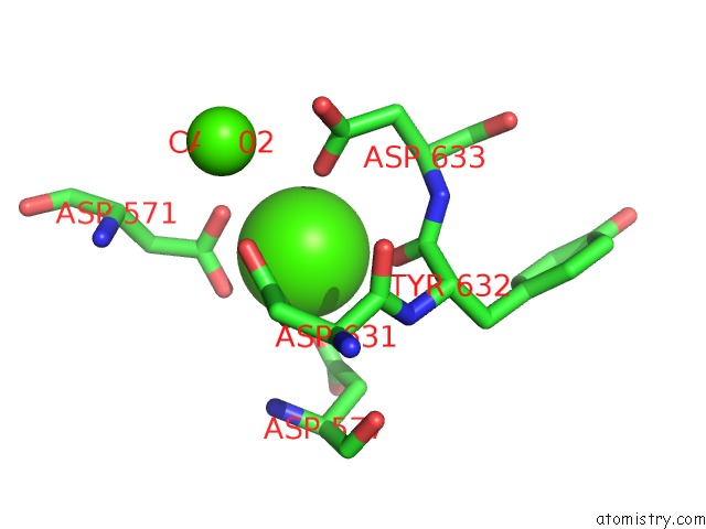
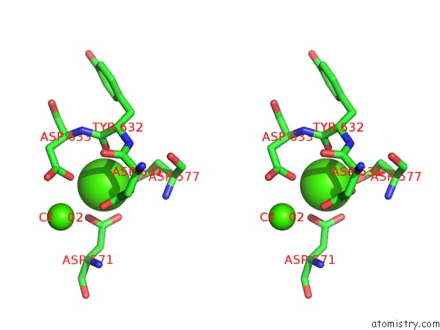
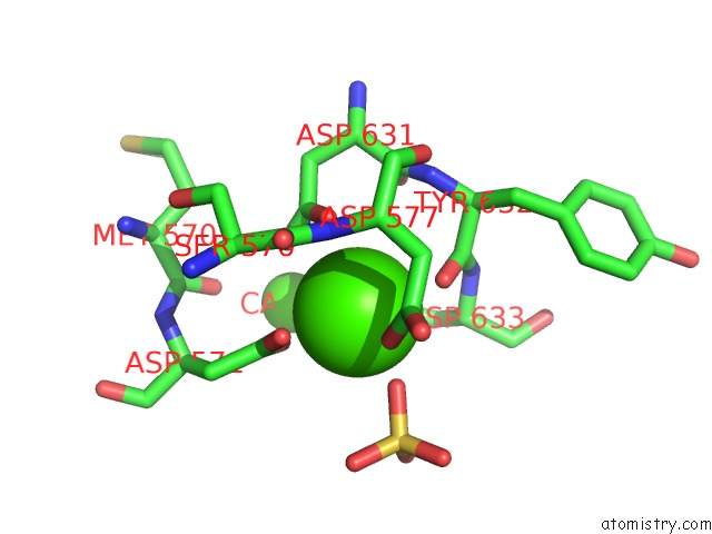
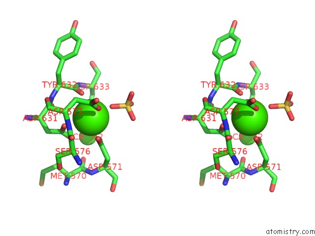
Calcium binding site 1 out of 6 in 5lob
Go back to
Calcium binding site 1 out
of 6 in the Structure of the CA2+-Bound RABPHILIN3A C2B- SNAP25 Complex (C2 Space Group)
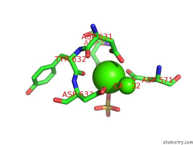
Mono view
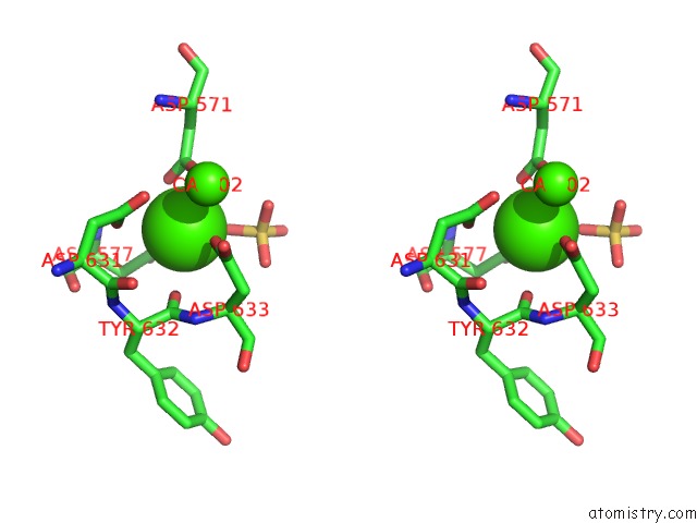
Stereo pair view
Mono view
Stereo pair view
A full contact list of Calcium with other atoms in the Ca binding
site number 1 of Structure of the CA2+-Bound RABPHILIN3A C2B- SNAP25 Complex (C2 Space Group) within 5.0Å range:
|
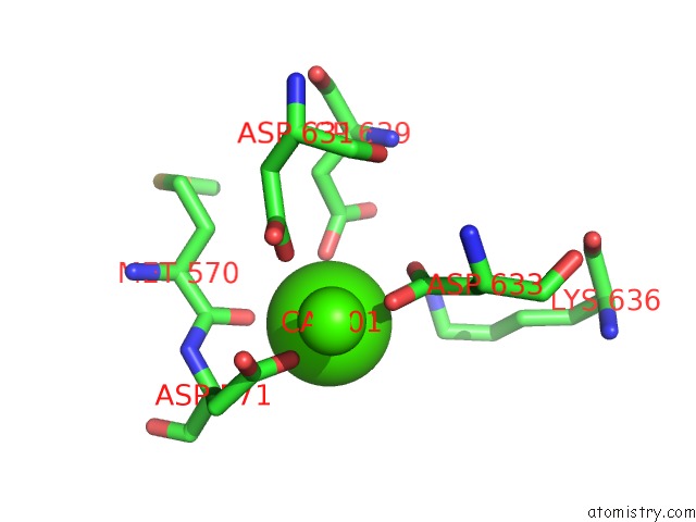
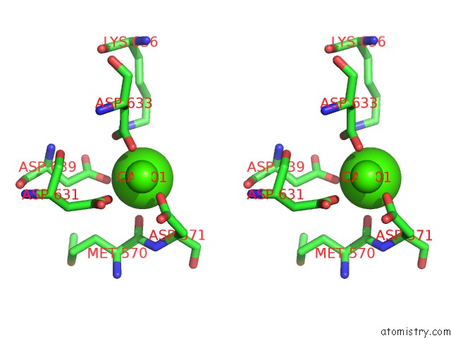
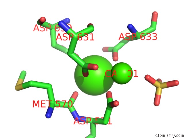
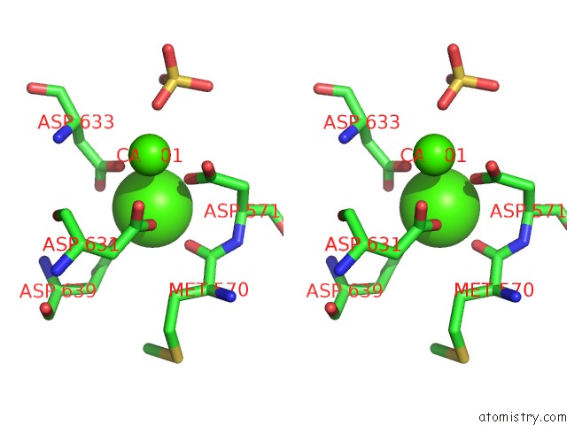
Calcium binding site 2 out of 6 in 5lob
Go back to
Calcium binding site 2 out
of 6 in the Structure of the CA2+-Bound RABPHILIN3A C2B- SNAP25 Complex (C2 Space Group)
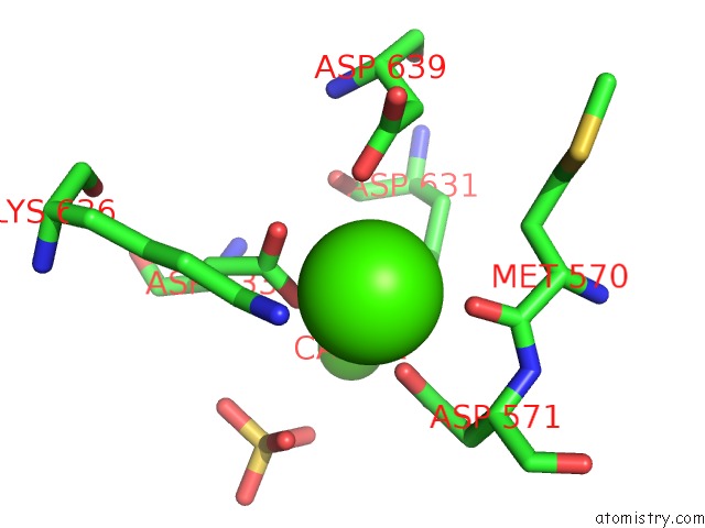
Mono view
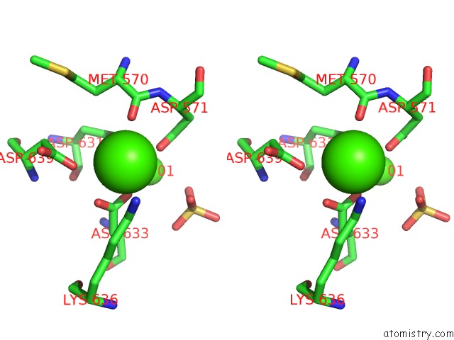
Stereo pair view
Mono view
Stereo pair view
A full contact list of Calcium with other atoms in the Ca binding
site number 2 of Structure of the CA2+-Bound RABPHILIN3A C2B- SNAP25 Complex (C2 Space Group) within 5.0Å range:
|
Calcium binding site 3 out of 6 in 5lob
Go back to
Calcium binding site 3 out
of 6 in the Structure of the CA2+-Bound RABPHILIN3A C2B- SNAP25 Complex (C2 Space Group)
Mono view
Stereo pair view
Mono view
Stereo pair view
A full contact list of Calcium with other atoms in the Ca binding
site number 3 of Structure of the CA2+-Bound RABPHILIN3A C2B- SNAP25 Complex (C2 Space Group) within 5.0Å range:
|
Calcium binding site 4 out of 6 in 5lob
Go back to
Calcium binding site 4 out
of 6 in the Structure of the CA2+-Bound RABPHILIN3A C2B- SNAP25 Complex (C2 Space Group)
Mono view
Stereo pair view
Mono view
Stereo pair view
A full contact list of Calcium with other atoms in the Ca binding
site number 4 of Structure of the CA2+-Bound RABPHILIN3A C2B- SNAP25 Complex (C2 Space Group) within 5.0Å range:
|
Calcium binding site 5 out of 6 in 5lob
Go back to
Calcium binding site 5 out
of 6 in the Structure of the CA2+-Bound RABPHILIN3A C2B- SNAP25 Complex (C2 Space Group)
Mono view
Stereo pair view
Mono view
Stereo pair view
A full contact list of Calcium with other atoms in the Ca binding
site number 5 of Structure of the CA2+-Bound RABPHILIN3A C2B- SNAP25 Complex (C2 Space Group) within 5.0Å range:
|
Calcium binding site 6 out of 6 in 5lob
Go back to
Calcium binding site 6 out
of 6 in the Structure of the CA2+-Bound RABPHILIN3A C2B- SNAP25 Complex (C2 Space Group)
Mono view
Stereo pair view
Mono view
Stereo pair view
A full contact list of Calcium with other atoms in the Ca binding
site number 6 of Structure of the CA2+-Bound RABPHILIN3A C2B- SNAP25 Complex (C2 Space Group) within 5.0Å range:
|
Reference:
C.Ferrer-Orta,
M.D.Perez-Sanchez,
T.Coronado-Parra,
C.Silva,
D.Lopez-Martinez,
J.Baltanas-Copado,
J.C.Gomez-Fernandez,
S.Corbalan-Garcia,
N.Verdaguer.
Structural Characterization of the Rabphilin-3A-SNAP25 Interaction. Proc. Natl. Acad. Sci. V. 114 E5343 2017U.S.A..
ISSN: ESSN 1091-6490
PubMed: 28634303
DOI: 10.1073/PNAS.1702542114
Page generated: Mon Jul 15 07:41:43 2024
ISSN: ESSN 1091-6490
PubMed: 28634303
DOI: 10.1073/PNAS.1702542114
Last articles
Zn in 9J0NZn in 9J0O
Zn in 9J0P
Zn in 9FJX
Zn in 9EKB
Zn in 9C0F
Zn in 9CAH
Zn in 9CH0
Zn in 9CH3
Zn in 9CH1