Calcium »
PDB 5lif-5m2o »
5lq2 »
Calcium in PDB 5lq2: Crystal Structure of TYR24 Phosphorylated Annexin A2 at 3.4 A Resolution
Protein crystallography data
The structure of Crystal Structure of TYR24 Phosphorylated Annexin A2 at 3.4 A Resolution, PDB code: 5lq2
was solved by
P.Ecsedi,
G.Gogl,
B.Kiss,
L.Nyitray,
with X-Ray Crystallography technique. A brief refinement statistics is given in the table below:
| Resolution Low / High (Å) | 86.51 / 3.40 |
| Space group | P 21 21 21 |
| Cell size a, b, c (Å), α, β, γ (°) | 61.370, 99.200, 173.020, 90.00, 90.00, 90.00 |
| R / Rfree (%) | 19.7 / 23.4 |
Calcium Binding Sites:
The binding sites of Calcium atom in the Crystal Structure of TYR24 Phosphorylated Annexin A2 at 3.4 A Resolution
(pdb code 5lq2). This binding sites where shown within
5.0 Angstroms radius around Calcium atom.
In total 10 binding sites of Calcium where determined in the Crystal Structure of TYR24 Phosphorylated Annexin A2 at 3.4 A Resolution, PDB code: 5lq2:
Jump to Calcium binding site number: 1; 2; 3; 4; 5; 6; 7; 8; 9; 10;
In total 10 binding sites of Calcium where determined in the Crystal Structure of TYR24 Phosphorylated Annexin A2 at 3.4 A Resolution, PDB code: 5lq2:
Jump to Calcium binding site number: 1; 2; 3; 4; 5; 6; 7; 8; 9; 10;
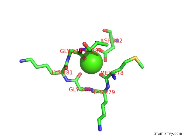
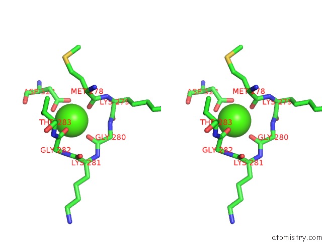
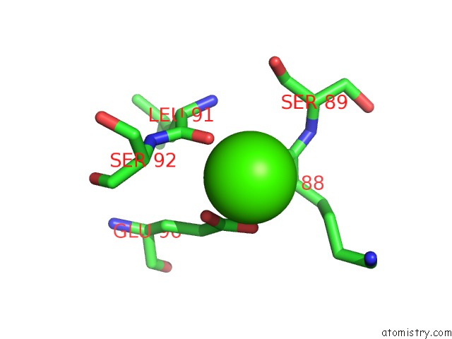
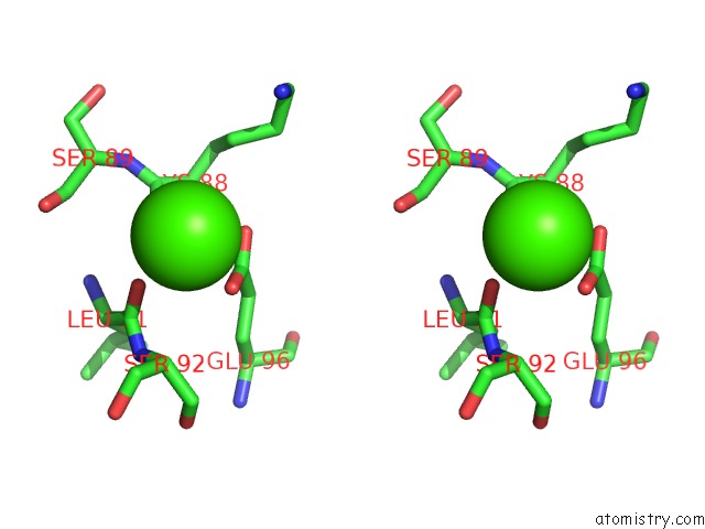
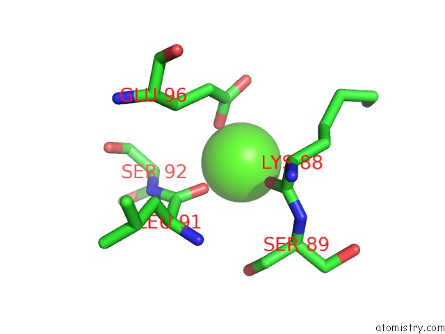
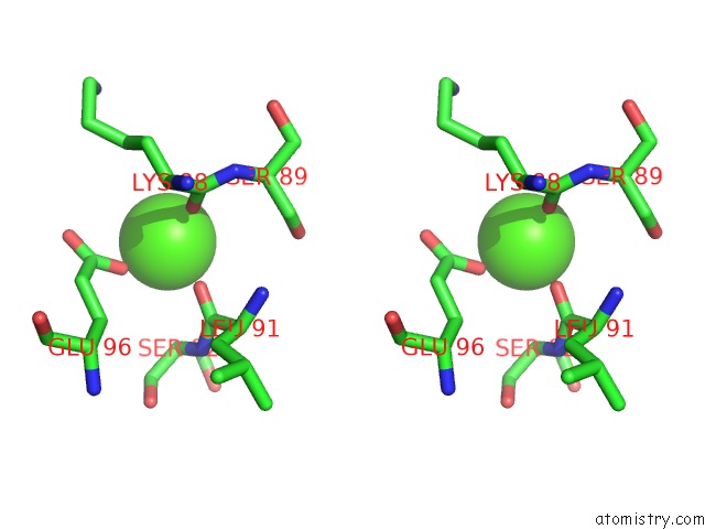
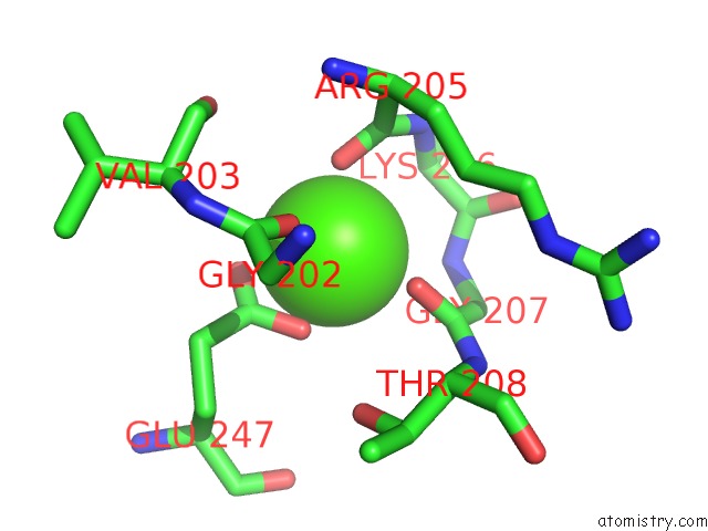
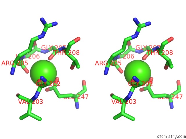
Calcium binding site 1 out of 10 in 5lq2
Go back to
Calcium binding site 1 out
of 10 in the Crystal Structure of TYR24 Phosphorylated Annexin A2 at 3.4 A Resolution
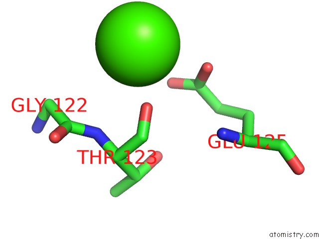
Mono view
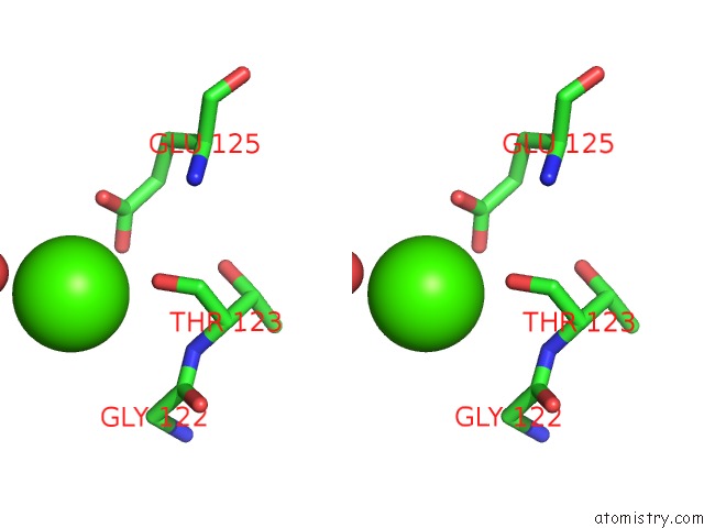
Stereo pair view
Mono view
Stereo pair view
A full contact list of Calcium with other atoms in the Ca binding
site number 1 of Crystal Structure of TYR24 Phosphorylated Annexin A2 at 3.4 A Resolution within 5.0Å range:
|
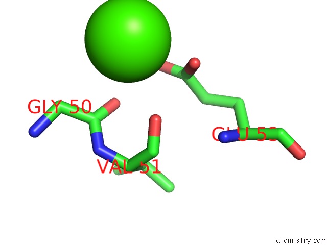
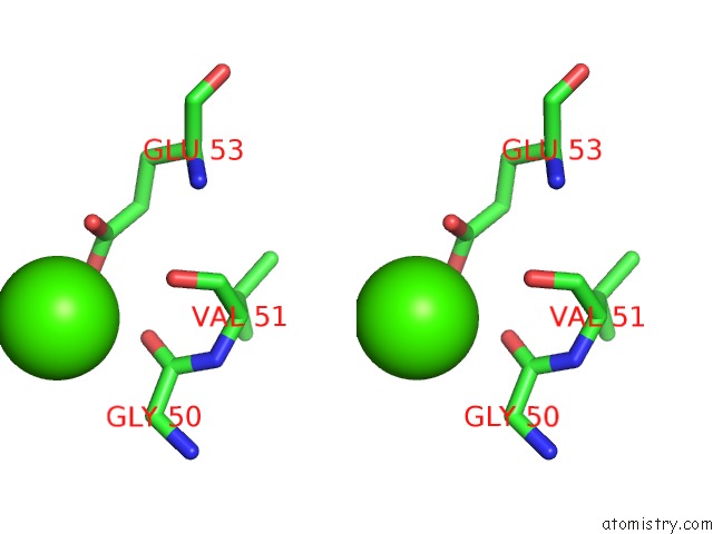
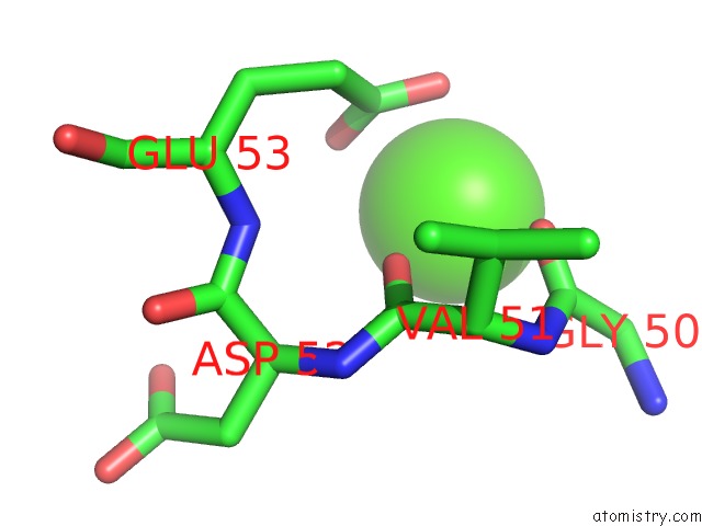
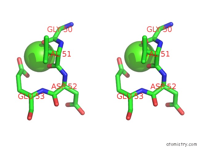
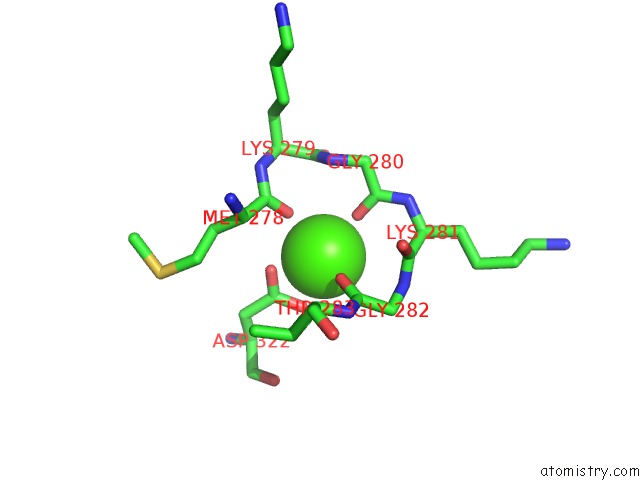
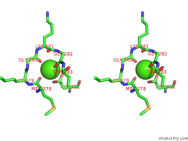
Calcium binding site 2 out of 10 in 5lq2
Go back to
Calcium binding site 2 out
of 10 in the Crystal Structure of TYR24 Phosphorylated Annexin A2 at 3.4 A Resolution
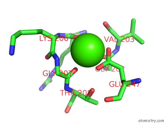
Mono view
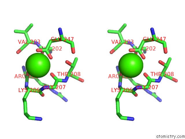
Stereo pair view
Mono view
Stereo pair view
A full contact list of Calcium with other atoms in the Ca binding
site number 2 of Crystal Structure of TYR24 Phosphorylated Annexin A2 at 3.4 A Resolution within 5.0Å range:
|
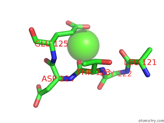
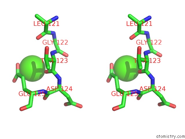
Calcium binding site 3 out of 10 in 5lq2
Go back to
Calcium binding site 3 out
of 10 in the Crystal Structure of TYR24 Phosphorylated Annexin A2 at 3.4 A Resolution
Mono view
Stereo pair view
Mono view
Stereo pair view
A full contact list of Calcium with other atoms in the Ca binding
site number 3 of Crystal Structure of TYR24 Phosphorylated Annexin A2 at 3.4 A Resolution within 5.0Å range:
|
Calcium binding site 4 out of 10 in 5lq2
Go back to
Calcium binding site 4 out
of 10 in the Crystal Structure of TYR24 Phosphorylated Annexin A2 at 3.4 A Resolution
Mono view
Stereo pair view
Mono view
Stereo pair view
A full contact list of Calcium with other atoms in the Ca binding
site number 4 of Crystal Structure of TYR24 Phosphorylated Annexin A2 at 3.4 A Resolution within 5.0Å range:
|
Calcium binding site 5 out of 10 in 5lq2
Go back to
Calcium binding site 5 out
of 10 in the Crystal Structure of TYR24 Phosphorylated Annexin A2 at 3.4 A Resolution
Mono view
Stereo pair view
Mono view
Stereo pair view
A full contact list of Calcium with other atoms in the Ca binding
site number 5 of Crystal Structure of TYR24 Phosphorylated Annexin A2 at 3.4 A Resolution within 5.0Å range:
|
Calcium binding site 6 out of 10 in 5lq2
Go back to
Calcium binding site 6 out
of 10 in the Crystal Structure of TYR24 Phosphorylated Annexin A2 at 3.4 A Resolution
Mono view
Stereo pair view
Mono view
Stereo pair view
A full contact list of Calcium with other atoms in the Ca binding
site number 6 of Crystal Structure of TYR24 Phosphorylated Annexin A2 at 3.4 A Resolution within 5.0Å range:
|
Calcium binding site 7 out of 10 in 5lq2
Go back to
Calcium binding site 7 out
of 10 in the Crystal Structure of TYR24 Phosphorylated Annexin A2 at 3.4 A Resolution
Mono view
Stereo pair view
Mono view
Stereo pair view
A full contact list of Calcium with other atoms in the Ca binding
site number 7 of Crystal Structure of TYR24 Phosphorylated Annexin A2 at 3.4 A Resolution within 5.0Å range:
|
Calcium binding site 8 out of 10 in 5lq2
Go back to
Calcium binding site 8 out
of 10 in the Crystal Structure of TYR24 Phosphorylated Annexin A2 at 3.4 A Resolution
Mono view
Stereo pair view
Mono view
Stereo pair view
A full contact list of Calcium with other atoms in the Ca binding
site number 8 of Crystal Structure of TYR24 Phosphorylated Annexin A2 at 3.4 A Resolution within 5.0Å range:
|
Calcium binding site 9 out of 10 in 5lq2
Go back to
Calcium binding site 9 out
of 10 in the Crystal Structure of TYR24 Phosphorylated Annexin A2 at 3.4 A Resolution
Mono view
Stereo pair view
Mono view
Stereo pair view
A full contact list of Calcium with other atoms in the Ca binding
site number 9 of Crystal Structure of TYR24 Phosphorylated Annexin A2 at 3.4 A Resolution within 5.0Å range:
|
Calcium binding site 10 out of 10 in 5lq2
Go back to
Calcium binding site 10 out
of 10 in the Crystal Structure of TYR24 Phosphorylated Annexin A2 at 3.4 A Resolution
Mono view
Stereo pair view
Mono view
Stereo pair view
A full contact list of Calcium with other atoms in the Ca binding
site number 10 of Crystal Structure of TYR24 Phosphorylated Annexin A2 at 3.4 A Resolution within 5.0Å range:
|
Reference:
P.Ecsedi,
B.Kiss,
G.Gogl,
L.Radnai,
L.Buday,
K.Koprivanacz,
K.Liliom,
I.Leveles,
B.Vertessy,
N.Jeszenoi,
C.Hetenyi,
G.Schlosser,
G.Katona,
L.Nyitray.
Regulation of the Equilibrium Between Closed and Open Conformations of Annexin A2 By N-Terminal Phosphorylation and S100A4-Binding. Structure V. 25 1195 2017.
ISSN: ISSN 1878-4186
PubMed: 28669632
DOI: 10.1016/J.STR.2017.06.001
Page generated: Mon Jul 15 07:45:17 2024
ISSN: ISSN 1878-4186
PubMed: 28669632
DOI: 10.1016/J.STR.2017.06.001
Last articles
Zn in 9J0NZn in 9J0O
Zn in 9J0P
Zn in 9FJX
Zn in 9EKB
Zn in 9C0F
Zn in 9CAH
Zn in 9CH0
Zn in 9CH3
Zn in 9CH1