Calcium »
PDB 5lif-5m2o »
5ls4 »
Calcium in PDB 5ls4: Mopeia Virus Exonuclease Domain Complexed with Calcium
Protein crystallography data
The structure of Mopeia Virus Exonuclease Domain Complexed with Calcium, PDB code: 5ls4
was solved by
E.L.Yekwa,
B.Canard,
F.Ferron,
with X-Ray Crystallography technique. A brief refinement statistics is given in the table below:
| Resolution Low / High (Å) | 47.52 / 1.47 |
| Space group | C 1 2 1 |
| Cell size a, b, c (Å), α, β, γ (°) | 131.962, 37.913, 48.986, 90.00, 104.07, 90.00 |
| R / Rfree (%) | 17 / 19.7 |
Other elements in 5ls4:
The structure of Mopeia Virus Exonuclease Domain Complexed with Calcium also contains other interesting chemical elements:
| Zinc | (Zn) | 1 atom |
Calcium Binding Sites:
The binding sites of Calcium atom in the Mopeia Virus Exonuclease Domain Complexed with Calcium
(pdb code 5ls4). This binding sites where shown within
5.0 Angstroms radius around Calcium atom.
In total only one binding site of Calcium was determined in the Mopeia Virus Exonuclease Domain Complexed with Calcium, PDB code: 5ls4:
In total only one binding site of Calcium was determined in the Mopeia Virus Exonuclease Domain Complexed with Calcium, PDB code: 5ls4:
Calcium binding site 1 out of 1 in 5ls4
Go back to
Calcium binding site 1 out
of 1 in the Mopeia Virus Exonuclease Domain Complexed with Calcium
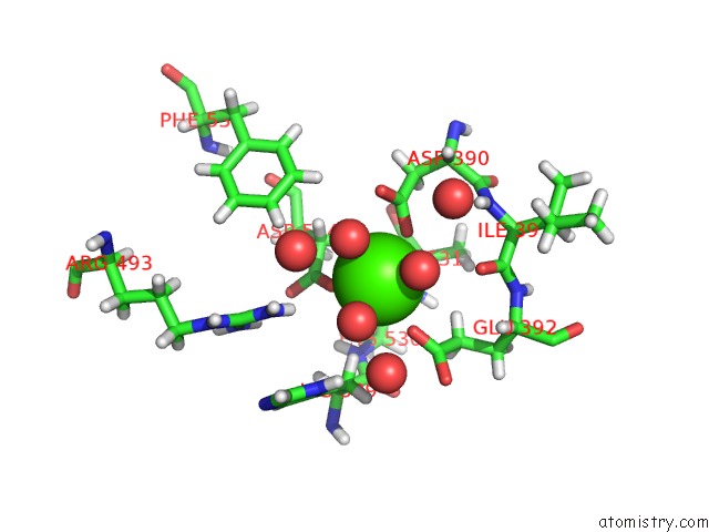
Mono view
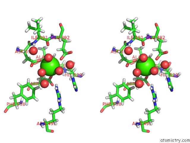
Stereo pair view
Mono view
Stereo pair view
A full contact list of Calcium with other atoms in the Ca binding
site number 1 of Mopeia Virus Exonuclease Domain Complexed with Calcium within 5.0Å range:
|
Reference:
E.Yekwa,
J.Khourieh,
B.Canard,
N.Papageorgiou,
F.Ferron.
Activity Inhibition and Crystal Polymorphism Induced By Active-Site Metal Swapping. Acta Crystallogr D Struct V. 73 641 2017BIOL.
ISSN: ISSN 2059-7983
PubMed: 28777079
DOI: 10.1107/S205979831700866X
Page generated: Wed Jul 9 07:59:48 2025
ISSN: ISSN 2059-7983
PubMed: 28777079
DOI: 10.1107/S205979831700866X
Last articles
Ca in 7G2QCa in 7G2P
Ca in 7G2N
Ca in 7G2O
Ca in 7G2M
Ca in 7G2L
Ca in 7G2K
Ca in 7G2J
Ca in 7G2I
Ca in 7G2G