Calcium »
PDB 5lif-5m2o »
5lul »
Calcium in PDB 5lul: Structure of A Triple Variant of Cutinase 2 From Thermobifida Cellulosilytica
Protein crystallography data
The structure of Structure of A Triple Variant of Cutinase 2 From Thermobifida Cellulosilytica, PDB code: 5lul
was solved by
A.Hromic,
A.Lyskowski,
K.Gruber,
with X-Ray Crystallography technique. A brief refinement statistics is given in the table below:
| Resolution Low / High (Å) | 44.53 / 1.90 |
| Space group | P 3 2 1 |
| Cell size a, b, c (Å), α, β, γ (°) | 110.438, 110.438, 75.302, 90.00, 90.00, 120.00 |
| R / Rfree (%) | 15.9 / 19.4 |
Other elements in 5lul:
The structure of Structure of A Triple Variant of Cutinase 2 From Thermobifida Cellulosilytica also contains other interesting chemical elements:
| Chlorine | (Cl) | 1 atom |
Calcium Binding Sites:
The binding sites of Calcium atom in the Structure of A Triple Variant of Cutinase 2 From Thermobifida Cellulosilytica
(pdb code 5lul). This binding sites where shown within
5.0 Angstroms radius around Calcium atom.
In total only one binding site of Calcium was determined in the Structure of A Triple Variant of Cutinase 2 From Thermobifida Cellulosilytica, PDB code: 5lul:
In total only one binding site of Calcium was determined in the Structure of A Triple Variant of Cutinase 2 From Thermobifida Cellulosilytica, PDB code: 5lul:
Calcium binding site 1 out of 1 in 5lul
Go back to
Calcium binding site 1 out
of 1 in the Structure of A Triple Variant of Cutinase 2 From Thermobifida Cellulosilytica
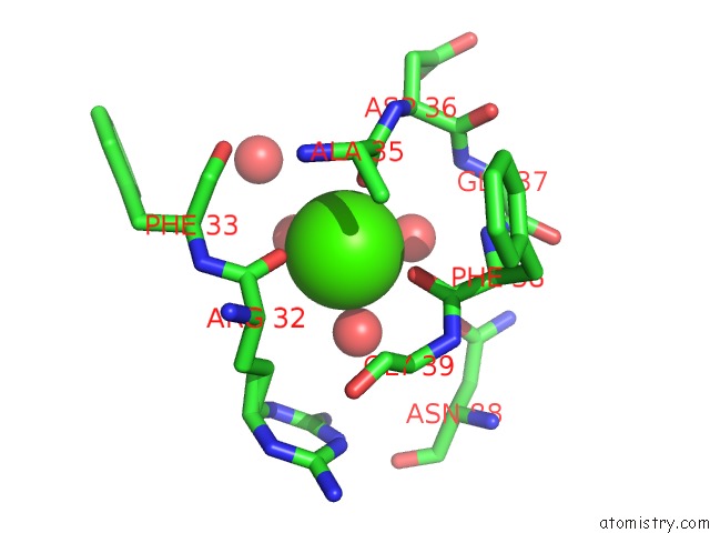
Mono view
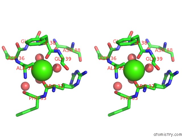
Stereo pair view
Mono view
Stereo pair view
A full contact list of Calcium with other atoms in the Ca binding
site number 1 of Structure of A Triple Variant of Cutinase 2 From Thermobifida Cellulosilytica within 5.0Å range:
|
Reference:
D.Ribitsch,
A.Hromic,
S.Zitzenbacher,
B.Zartl,
C.Gamerith,
A.Pellis,
A.Jungbauer,
A.Yskowski,
G.Steinkellner,
K.Gruber,
R.Tscheliessnig,
E.Herrero Acero,
G.M.Guebitz.
Small Cause, Large Effect: Structural Characterization of Cutinases From Thermobifida Cellulosilytica. Biotechnol. Bioeng. V. 114 2481 2017.
ISSN: ESSN 1097-0290
PubMed: 28671263
DOI: 10.1002/BIT.26372
Page generated: Mon Jul 15 07:48:20 2024
ISSN: ESSN 1097-0290
PubMed: 28671263
DOI: 10.1002/BIT.26372
Last articles
Zn in 9MJ5Zn in 9HNW
Zn in 9G0L
Zn in 9FNE
Zn in 9DZN
Zn in 9E0I
Zn in 9D32
Zn in 9DAK
Zn in 8ZXC
Zn in 8ZUF