Calcium »
PDB 5lif-5m2o »
5lxs »
Calcium in PDB 5lxs: Tubulin-Ks-1-199-32 Complex
Protein crystallography data
The structure of Tubulin-Ks-1-199-32 Complex, PDB code: 5lxs
was solved by
A.E.Prota,
M.O.Steinmetz,
with X-Ray Crystallography technique. A brief refinement statistics is given in the table below:
| Resolution Low / High (Å) | 72.23 / 2.20 |
| Space group | P 21 21 21 |
| Cell size a, b, c (Å), α, β, γ (°) | 104.580, 157.800, 179.440, 90.00, 90.00, 90.00 |
| R / Rfree (%) | 18.7 / 22.7 |
Other elements in 5lxs:
The structure of Tubulin-Ks-1-199-32 Complex also contains other interesting chemical elements:
| Magnesium | (Mg) | 5 atoms |
Calcium Binding Sites:
The binding sites of Calcium atom in the Tubulin-Ks-1-199-32 Complex
(pdb code 5lxs). This binding sites where shown within
5.0 Angstroms radius around Calcium atom.
In total 4 binding sites of Calcium where determined in the Tubulin-Ks-1-199-32 Complex, PDB code: 5lxs:
Jump to Calcium binding site number: 1; 2; 3; 4;
In total 4 binding sites of Calcium where determined in the Tubulin-Ks-1-199-32 Complex, PDB code: 5lxs:
Jump to Calcium binding site number: 1; 2; 3; 4;
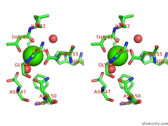
Calcium binding site 1 out of 4 in 5lxs
Go back to
Calcium binding site 1 out
of 4 in the Tubulin-Ks-1-199-32 Complex

Mono view

Stereo pair view
Mono view
Stereo pair view
A full contact list of Calcium with other atoms in the Ca binding
site number 1 of Tubulin-Ks-1-199-32 Complex within 5.0Å range:
|
Calcium binding site 2 out of 4 in 5lxs
Go back to
Calcium binding site 2 out
of 4 in the Tubulin-Ks-1-199-32 Complex

Mono view

Stereo pair view
Mono view
Stereo pair view
A full contact list of Calcium with other atoms in the Ca binding
site number 2 of Tubulin-Ks-1-199-32 Complex within 5.0Å range:
|
Calcium binding site 3 out of 4 in 5lxs
Go back to
Calcium binding site 3 out
of 4 in the Tubulin-Ks-1-199-32 Complex

Mono view
Stereo pair view
Mono view
Stereo pair view
A full contact list of Calcium with other atoms in the Ca binding
site number 3 of Tubulin-Ks-1-199-32 Complex within 5.0Å range:
|
Calcium binding site 4 out of 4 in 5lxs
Go back to
Calcium binding site 4 out
of 4 in the Tubulin-Ks-1-199-32 Complex

Mono view

Stereo pair view
Mono view
Stereo pair view
A full contact list of Calcium with other atoms in the Ca binding
site number 4 of Tubulin-Ks-1-199-32 Complex within 5.0Å range:
|
Reference:
A.E.Prota,
K.Bargsten,
M.Redondo-Horcajo,
A.B.Smith,
C.H.Yang,
H.M.Mcdaid,
I.Paterson,
S.B.Horwitz,
J.Fernando Diaz,
M.O.Steinmetz.
Structural Basis of Microtubule Stabilization By Discodermolide. Chembiochem V. 18 905 2017.
ISSN: ESSN 1439-7633
PubMed: 28207984
DOI: 10.1002/CBIC.201600696
Page generated: Wed Jul 9 08:01:26 2025
ISSN: ESSN 1439-7633
PubMed: 28207984
DOI: 10.1002/CBIC.201600696
Last articles
Cl in 5LANCl in 5LAG
Cl in 5LAF
Cl in 5LA8
Cl in 5LA5
Cl in 5L9J
Cl in 5L9Z
Cl in 5L7I
Cl in 5L8D
Cl in 5L7U