Calcium »
PDB 5m2s-5mi4 »
5m8e »
Calcium in PDB 5m8e: Crystal Structure of A GH43 Arabonofuranosidase From Weissella Sp. Strain 142
Enzymatic activity of Crystal Structure of A GH43 Arabonofuranosidase From Weissella Sp. Strain 142
All present enzymatic activity of Crystal Structure of A GH43 Arabonofuranosidase From Weissella Sp. Strain 142:
3.2.1.55;
3.2.1.55;
Protein crystallography data
The structure of Crystal Structure of A GH43 Arabonofuranosidase From Weissella Sp. Strain 142, PDB code: 5m8e
was solved by
J.Linares-Pasten,
D.T.Logan,
E.Nordberg Karlsson,
with X-Ray Crystallography technique. A brief refinement statistics is given in the table below:
| Resolution Low / High (Å) | 29.70 / 2.00 |
| Space group | P 1 21 1 |
| Cell size a, b, c (Å), α, β, γ (°) | 59.880, 71.930, 79.510, 90.00, 101.93, 90.00 |
| R / Rfree (%) | 17.2 / 21.6 |
Calcium Binding Sites:
The binding sites of Calcium atom in the Crystal Structure of A GH43 Arabonofuranosidase From Weissella Sp. Strain 142
(pdb code 5m8e). This binding sites where shown within
5.0 Angstroms radius around Calcium atom.
In total 2 binding sites of Calcium where determined in the Crystal Structure of A GH43 Arabonofuranosidase From Weissella Sp. Strain 142, PDB code: 5m8e:
Jump to Calcium binding site number: 1; 2;
In total 2 binding sites of Calcium where determined in the Crystal Structure of A GH43 Arabonofuranosidase From Weissella Sp. Strain 142, PDB code: 5m8e:
Jump to Calcium binding site number: 1; 2;
Calcium binding site 1 out of 2 in 5m8e
Go back to
Calcium binding site 1 out
of 2 in the Crystal Structure of A GH43 Arabonofuranosidase From Weissella Sp. Strain 142
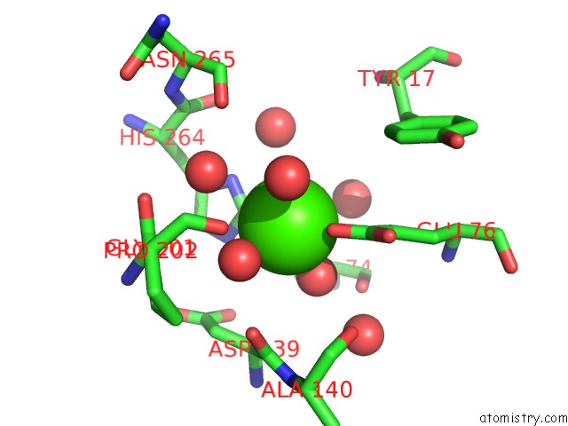
Mono view
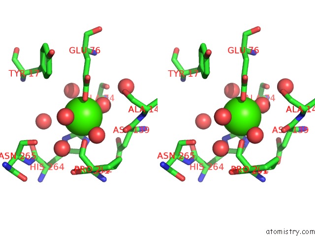
Stereo pair view
Mono view
Stereo pair view
A full contact list of Calcium with other atoms in the Ca binding
site number 1 of Crystal Structure of A GH43 Arabonofuranosidase From Weissella Sp. Strain 142 within 5.0Å range:
|
Calcium binding site 2 out of 2 in 5m8e
Go back to
Calcium binding site 2 out
of 2 in the Crystal Structure of A GH43 Arabonofuranosidase From Weissella Sp. Strain 142
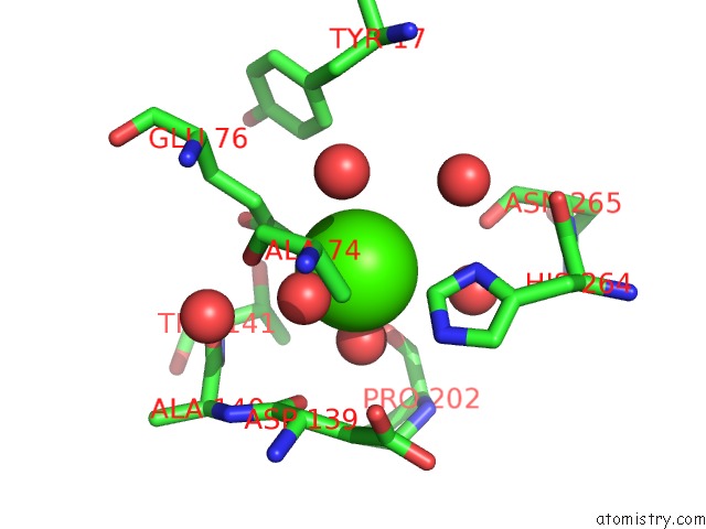
Mono view
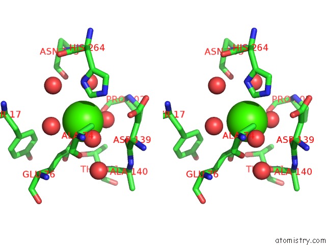
Stereo pair view
Mono view
Stereo pair view
A full contact list of Calcium with other atoms in the Ca binding
site number 2 of Crystal Structure of A GH43 Arabonofuranosidase From Weissella Sp. Strain 142 within 5.0Å range:
|
Reference:
J.A.Linares-Pasten,
P.Falck,
K.Albasri,
S.Kjellstrom,
P.Adlercreutz,
D.T.Logan,
E.N.Karlsson.
Three-Dimensional Structures and Functional Studies of Two GH43 Arabinofuranosidases From Weissella Sp. Strain 142 and Lactobacillus Brevis. Febs J. V. 284 2019 2017.
ISSN: ISSN 1742-4658
PubMed: 28485897
DOI: 10.1111/FEBS.14101
Page generated: Mon Jul 15 07:59:29 2024
ISSN: ISSN 1742-4658
PubMed: 28485897
DOI: 10.1111/FEBS.14101
Last articles
Zn in 9J0NZn in 9J0O
Zn in 9J0P
Zn in 9FJX
Zn in 9EKB
Zn in 9C0F
Zn in 9CAH
Zn in 9CH0
Zn in 9CH3
Zn in 9CH1