Calcium »
PDB 5m2s-5mi4 »
5m9w »
Calcium in PDB 5m9w: Experimental Mad Phased Structure of Thermolysin in Complex with Inhibitor JC65.
Enzymatic activity of Experimental Mad Phased Structure of Thermolysin in Complex with Inhibitor JC65.
All present enzymatic activity of Experimental Mad Phased Structure of Thermolysin in Complex with Inhibitor JC65.:
3.4.24.27;
3.4.24.27;
Protein crystallography data
The structure of Experimental Mad Phased Structure of Thermolysin in Complex with Inhibitor JC65., PDB code: 5m9w
was solved by
S.G.Krimmer,
J.Cramer,
A.Heine,
G.Klebe,
with X-Ray Crystallography technique. A brief refinement statistics is given in the table below:
| Resolution Low / High (Å) | 43.60 / 1.21 |
| Space group | P 61 2 2 |
| Cell size a, b, c (Å), α, β, γ (°) | 92.516, 92.516, 130.503, 90.00, 90.00, 120.00 |
| R / Rfree (%) | 10.7 / 12.7 |
Other elements in 5m9w:
The structure of Experimental Mad Phased Structure of Thermolysin in Complex with Inhibitor JC65. also contains other interesting chemical elements:
| Zinc | (Zn) | 1 atom |
Calcium Binding Sites:
The binding sites of Calcium atom in the Experimental Mad Phased Structure of Thermolysin in Complex with Inhibitor JC65.
(pdb code 5m9w). This binding sites where shown within
5.0 Angstroms radius around Calcium atom.
In total 4 binding sites of Calcium where determined in the Experimental Mad Phased Structure of Thermolysin in Complex with Inhibitor JC65., PDB code: 5m9w:
Jump to Calcium binding site number: 1; 2; 3; 4;
In total 4 binding sites of Calcium where determined in the Experimental Mad Phased Structure of Thermolysin in Complex with Inhibitor JC65., PDB code: 5m9w:
Jump to Calcium binding site number: 1; 2; 3; 4;
Calcium binding site 1 out of 4 in 5m9w
Go back to
Calcium binding site 1 out
of 4 in the Experimental Mad Phased Structure of Thermolysin in Complex with Inhibitor JC65.
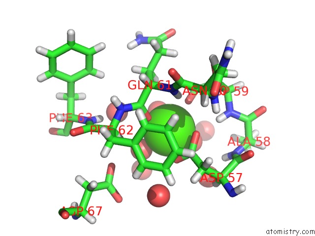
Mono view

Stereo pair view
Mono view
Stereo pair view
A full contact list of Calcium with other atoms in the Ca binding
site number 1 of Experimental Mad Phased Structure of Thermolysin in Complex with Inhibitor JC65. within 5.0Å range:
|
Calcium binding site 2 out of 4 in 5m9w
Go back to
Calcium binding site 2 out
of 4 in the Experimental Mad Phased Structure of Thermolysin in Complex with Inhibitor JC65.

Mono view
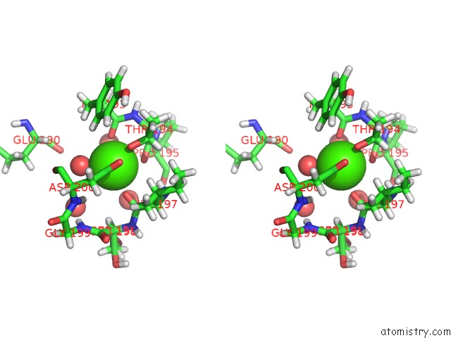
Stereo pair view
Mono view
Stereo pair view
A full contact list of Calcium with other atoms in the Ca binding
site number 2 of Experimental Mad Phased Structure of Thermolysin in Complex with Inhibitor JC65. within 5.0Å range:
|
Calcium binding site 3 out of 4 in 5m9w
Go back to
Calcium binding site 3 out
of 4 in the Experimental Mad Phased Structure of Thermolysin in Complex with Inhibitor JC65.

Mono view
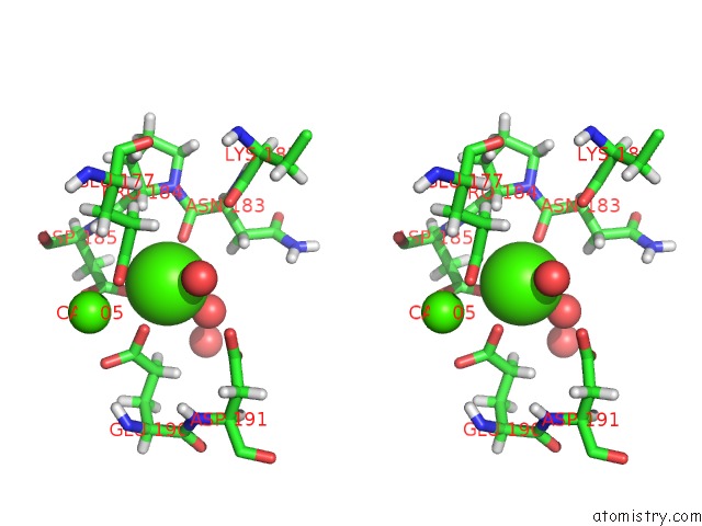
Stereo pair view
Mono view
Stereo pair view
A full contact list of Calcium with other atoms in the Ca binding
site number 3 of Experimental Mad Phased Structure of Thermolysin in Complex with Inhibitor JC65. within 5.0Å range:
|
Calcium binding site 4 out of 4 in 5m9w
Go back to
Calcium binding site 4 out
of 4 in the Experimental Mad Phased Structure of Thermolysin in Complex with Inhibitor JC65.
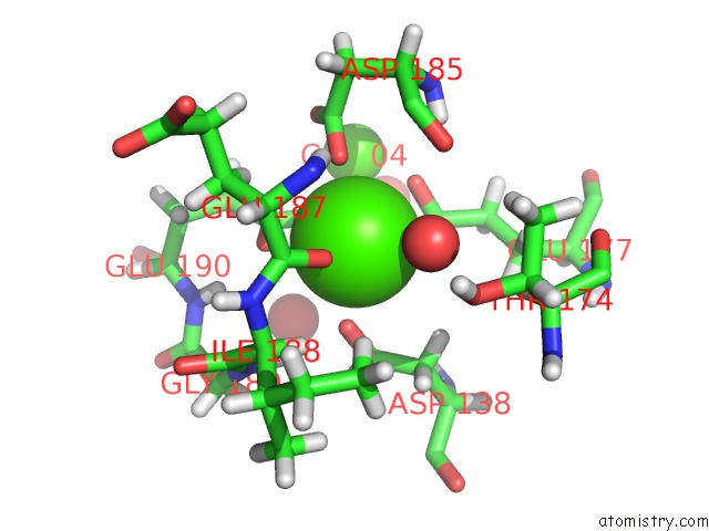
Mono view

Stereo pair view
Mono view
Stereo pair view
A full contact list of Calcium with other atoms in the Ca binding
site number 4 of Experimental Mad Phased Structure of Thermolysin in Complex with Inhibitor JC65. within 5.0Å range:
|
Reference:
S.G.Krimmer,
J.Cramer,
J.Schiebel,
A.Heine,
G.Klebe.
How Nothing Boosts Affinity: Hydrophobic Ligand Binding to the Virtually Vacated S1' Pocket of Thermolysin. J. Am. Chem. Soc. V. 139 10419 2017.
ISSN: ESSN 1520-5126
PubMed: 28696673
DOI: 10.1021/JACS.7B05028
Page generated: Wed Jul 9 08:07:53 2025
ISSN: ESSN 1520-5126
PubMed: 28696673
DOI: 10.1021/JACS.7B05028
Last articles
F in 4LW1F in 4LWU
F in 4LTS
F in 4LUD
F in 4LUV
F in 4LOQ
F in 4LQG
F in 4LPB
F in 4LOP
F in 4LP0