Calcium »
PDB 5mi5-5mop »
5mim »
Calcium in PDB 5mim: Xray Structure of Human Furin Bound with the 2,5-Dideoxystreptamine Derived Small Molecule Inhibitor 1N
Enzymatic activity of Xray Structure of Human Furin Bound with the 2,5-Dideoxystreptamine Derived Small Molecule Inhibitor 1N
All present enzymatic activity of Xray Structure of Human Furin Bound with the 2,5-Dideoxystreptamine Derived Small Molecule Inhibitor 1N:
3.4.21.75;
3.4.21.75;
Protein crystallography data
The structure of Xray Structure of Human Furin Bound with the 2,5-Dideoxystreptamine Derived Small Molecule Inhibitor 1N, PDB code: 5mim
was solved by
S.O.Dahms,
J.Guan-Sheng,
M.E.Than,
with X-Ray Crystallography technique. A brief refinement statistics is given in the table below:
| Resolution Low / High (Å) | 41.69 / 1.90 |
| Space group | P 65 2 2 |
| Cell size a, b, c (Å), α, β, γ (°) | 132.204, 132.204, 155.727, 90.00, 90.00, 120.00 |
| R / Rfree (%) | 16.5 / 18.4 |
Other elements in 5mim:
The structure of Xray Structure of Human Furin Bound with the 2,5-Dideoxystreptamine Derived Small Molecule Inhibitor 1N also contains other interesting chemical elements:
| Chlorine | (Cl) | 1 atom |
| Sodium | (Na) | 4 atoms |
Calcium Binding Sites:
The binding sites of Calcium atom in the Xray Structure of Human Furin Bound with the 2,5-Dideoxystreptamine Derived Small Molecule Inhibitor 1N
(pdb code 5mim). This binding sites where shown within
5.0 Angstroms radius around Calcium atom.
In total 3 binding sites of Calcium where determined in the Xray Structure of Human Furin Bound with the 2,5-Dideoxystreptamine Derived Small Molecule Inhibitor 1N, PDB code: 5mim:
Jump to Calcium binding site number: 1; 2; 3;
In total 3 binding sites of Calcium where determined in the Xray Structure of Human Furin Bound with the 2,5-Dideoxystreptamine Derived Small Molecule Inhibitor 1N, PDB code: 5mim:
Jump to Calcium binding site number: 1; 2; 3;
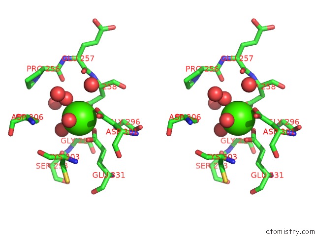
Calcium binding site 1 out of 3 in 5mim
Go back to
Calcium binding site 1 out
of 3 in the Xray Structure of Human Furin Bound with the 2,5-Dideoxystreptamine Derived Small Molecule Inhibitor 1N
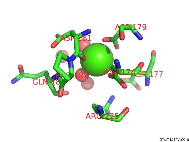
Mono view
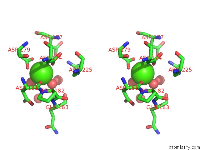
Stereo pair view
Mono view
Stereo pair view
A full contact list of Calcium with other atoms in the Ca binding
site number 1 of Xray Structure of Human Furin Bound with the 2,5-Dideoxystreptamine Derived Small Molecule Inhibitor 1N within 5.0Å range:
|
Calcium binding site 2 out of 3 in 5mim
Go back to
Calcium binding site 2 out
of 3 in the Xray Structure of Human Furin Bound with the 2,5-Dideoxystreptamine Derived Small Molecule Inhibitor 1N
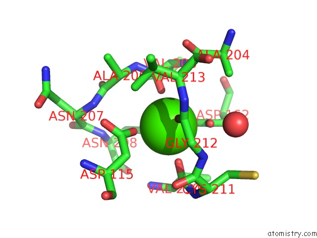
Mono view
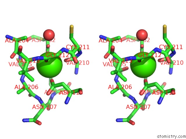
Stereo pair view
Mono view
Stereo pair view
A full contact list of Calcium with other atoms in the Ca binding
site number 2 of Xray Structure of Human Furin Bound with the 2,5-Dideoxystreptamine Derived Small Molecule Inhibitor 1N within 5.0Å range:
|
Calcium binding site 3 out of 3 in 5mim
Go back to
Calcium binding site 3 out
of 3 in the Xray Structure of Human Furin Bound with the 2,5-Dideoxystreptamine Derived Small Molecule Inhibitor 1N

Mono view
Stereo pair view
Mono view
Stereo pair view
A full contact list of Calcium with other atoms in the Ca binding
site number 3 of Xray Structure of Human Furin Bound with the 2,5-Dideoxystreptamine Derived Small Molecule Inhibitor 1N within 5.0Å range:
|
Reference:
S.O.Dahms,
G.S.Jiao,
M.E.Than.
Structural Studies Revealed Active Site Distortions of Human Furin By A Small Molecule Inhibitor. Acs Chem. Biol. V. 12 1211 2017.
ISSN: ESSN 1554-8937
PubMed: 28402100
DOI: 10.1021/ACSCHEMBIO.6B01110
Page generated: Mon Jul 15 08:26:20 2024
ISSN: ESSN 1554-8937
PubMed: 28402100
DOI: 10.1021/ACSCHEMBIO.6B01110
Last articles
Zn in 9MJ5Zn in 9HNW
Zn in 9G0L
Zn in 9FNE
Zn in 9DZN
Zn in 9E0I
Zn in 9D32
Zn in 9DAK
Zn in 8ZXC
Zn in 8ZUF