Calcium »
PDB 5mi5-5mop »
5mj7 »
Calcium in PDB 5mj7: Structure of the C. Elegans Nucleoside Hydrolase
Protein crystallography data
The structure of Structure of the C. Elegans Nucleoside Hydrolase, PDB code: 5mj7
was solved by
W.Versees,
R.K.Singh,
with X-Ray Crystallography technique. A brief refinement statistics is given in the table below:
| Resolution Low / High (Å) | 24.93 / 1.65 |
| Space group | P 41 21 2 |
| Cell size a, b, c (Å), α, β, γ (°) | 84.578, 84.578, 260.911, 90.00, 90.00, 90.00 |
| R / Rfree (%) | 13.7 / 15.4 |
Calcium Binding Sites:
The binding sites of Calcium atom in the Structure of the C. Elegans Nucleoside Hydrolase
(pdb code 5mj7). This binding sites where shown within
5.0 Angstroms radius around Calcium atom.
In total 2 binding sites of Calcium where determined in the Structure of the C. Elegans Nucleoside Hydrolase, PDB code: 5mj7:
Jump to Calcium binding site number: 1; 2;
In total 2 binding sites of Calcium where determined in the Structure of the C. Elegans Nucleoside Hydrolase, PDB code: 5mj7:
Jump to Calcium binding site number: 1; 2;
Calcium binding site 1 out of 2 in 5mj7
Go back to
Calcium binding site 1 out
of 2 in the Structure of the C. Elegans Nucleoside Hydrolase
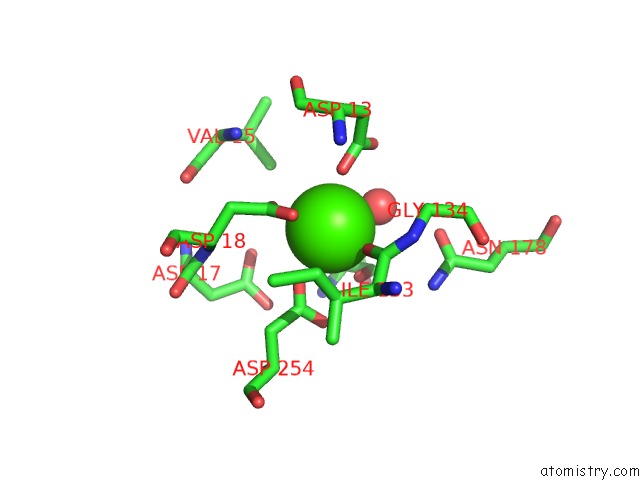
Mono view
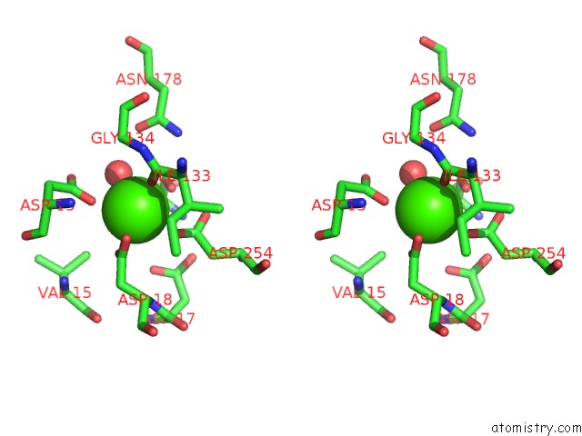
Stereo pair view
Mono view
Stereo pair view
A full contact list of Calcium with other atoms in the Ca binding
site number 1 of Structure of the C. Elegans Nucleoside Hydrolase within 5.0Å range:
|
Calcium binding site 2 out of 2 in 5mj7
Go back to
Calcium binding site 2 out
of 2 in the Structure of the C. Elegans Nucleoside Hydrolase
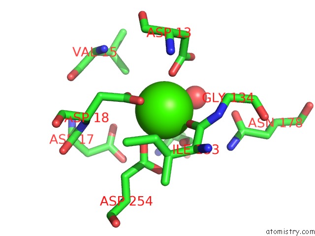
Mono view
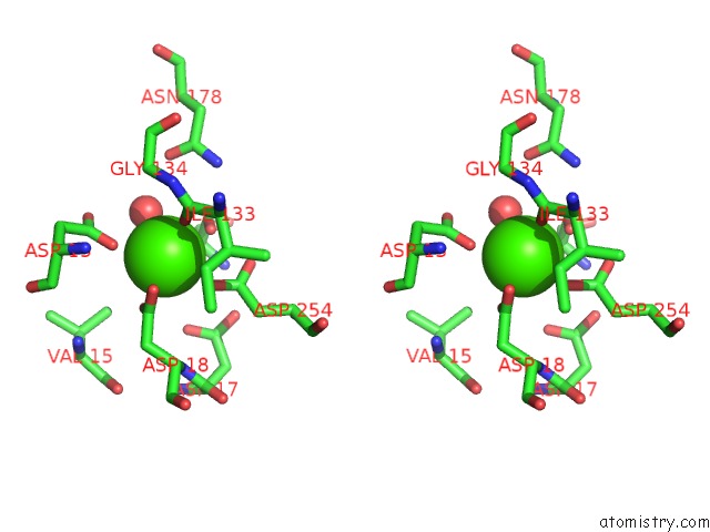
Stereo pair view
Mono view
Stereo pair view
A full contact list of Calcium with other atoms in the Ca binding
site number 2 of Structure of the C. Elegans Nucleoside Hydrolase within 5.0Å range:
|
Reference:
R.K.Singh,
J.Steyaert,
W.Versees.
Structural and Biochemical Characterization of the Nucleoside Hydrolase From C. Elegans Reveals the Role of Two Active Site Cysteine Residues in Catalysis. Protein Sci. V. 26 985 2017.
ISSN: ESSN 1469-896X
PubMed: 28218438
DOI: 10.1002/PRO.3141
Page generated: Mon Jul 15 08:26:20 2024
ISSN: ESSN 1469-896X
PubMed: 28218438
DOI: 10.1002/PRO.3141
Last articles
Zn in 9MJ5Zn in 9HNW
Zn in 9G0L
Zn in 9FNE
Zn in 9DZN
Zn in 9E0I
Zn in 9D32
Zn in 9DAK
Zn in 8ZXC
Zn in 8ZUF