Calcium »
PDB 5mi5-5mop »
5ml7 »
Calcium in PDB 5ml7: Crystal Structure of L1-Stalk Fragment of 23S Rrna From Haloarcula Marismortui
Protein crystallography data
The structure of Crystal Structure of L1-Stalk Fragment of 23S Rrna From Haloarcula Marismortui, PDB code: 5ml7
was solved by
A.G.Gabdulkhakov,
T.V.Tishchenko,
N.A.Nevskaya,
S.V.Nikonov,
with X-Ray Crystallography technique. A brief refinement statistics is given in the table below:
| Resolution Low / High (Å) | 30.00 / 3.30 |
| Space group | P 41 21 2 |
| Cell size a, b, c (Å), α, β, γ (°) | 136.836, 136.836, 70.110, 90.00, 90.00, 90.00 |
| R / Rfree (%) | 22.6 / 29.1 |
Other elements in 5ml7:
The structure of Crystal Structure of L1-Stalk Fragment of 23S Rrna From Haloarcula Marismortui also contains other interesting chemical elements:
| Magnesium | (Mg) | 8 atoms |
Calcium Binding Sites:
The binding sites of Calcium atom in the Crystal Structure of L1-Stalk Fragment of 23S Rrna From Haloarcula Marismortui
(pdb code 5ml7). This binding sites where shown within
5.0 Angstroms radius around Calcium atom.
In total only one binding site of Calcium was determined in the Crystal Structure of L1-Stalk Fragment of 23S Rrna From Haloarcula Marismortui, PDB code: 5ml7:
In total only one binding site of Calcium was determined in the Crystal Structure of L1-Stalk Fragment of 23S Rrna From Haloarcula Marismortui, PDB code: 5ml7:
Calcium binding site 1 out of 1 in 5ml7
Go back to
Calcium binding site 1 out
of 1 in the Crystal Structure of L1-Stalk Fragment of 23S Rrna From Haloarcula Marismortui
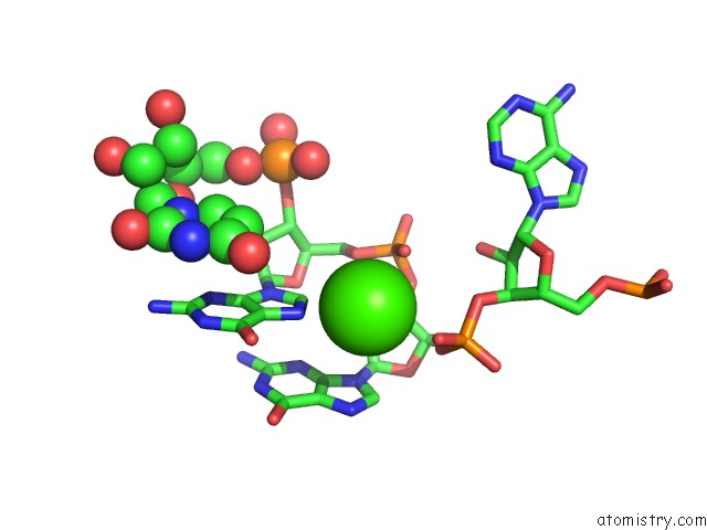
Mono view
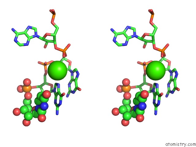
Stereo pair view
Mono view
Stereo pair view
A full contact list of Calcium with other atoms in the Ca binding
site number 1 of Crystal Structure of L1-Stalk Fragment of 23S Rrna From Haloarcula Marismortui within 5.0Å range:
|
Reference:
A.G.Gabdulkhakov,
T.V.Tishchenko,
A.Mikhaylina,
M.Garber,
N.A.Nevskaya,
S.V.Nikonov.
Crystal Structure of the 23S Rrna Fragment Specific to R-Protein L1 and Designed Model of the Ribosomal L1 Stalk From Haloarcula Marismortui Crystals 2017.
ISSN: ESSN 2073-4352
DOI: 10.3390/CRYST7020037
Page generated: Mon Jul 15 08:28:10 2024
ISSN: ESSN 2073-4352
DOI: 10.3390/CRYST7020037
Last articles
Zn in 9J0NZn in 9J0O
Zn in 9J0P
Zn in 9FJX
Zn in 9EKB
Zn in 9C0F
Zn in 9CAH
Zn in 9CH0
Zn in 9CH3
Zn in 9CH1