Calcium »
PDB 5mi5-5mop »
5mnf »
Calcium in PDB 5mnf: Cationic Trypsin in Its Apo Form (Deuterated Sample at 295 K)
Enzymatic activity of Cationic Trypsin in Its Apo Form (Deuterated Sample at 295 K)
All present enzymatic activity of Cationic Trypsin in Its Apo Form (Deuterated Sample at 295 K):
3.4.21.4;
3.4.21.4;
Protein crystallography data
The structure of Cationic Trypsin in Its Apo Form (Deuterated Sample at 295 K), PDB code: 5mnf
was solved by
J.Schiebel,
A.Heine,
G.Klebe,
with X-Ray Crystallography technique. A brief refinement statistics is given in the table below:
| Resolution Low / High (Å) | 19.23 / 0.99 |
| Space group | P 21 21 21 |
| Cell size a, b, c (Å), α, β, γ (°) | 54.999, 58.623, 67.565, 90.00, 90.00, 90.00 |
| R / Rfree (%) | 9.6 / 10.7 |
Calcium Binding Sites:
The binding sites of Calcium atom in the Cationic Trypsin in Its Apo Form (Deuterated Sample at 295 K)
(pdb code 5mnf). This binding sites where shown within
5.0 Angstroms radius around Calcium atom.
In total only one binding site of Calcium was determined in the Cationic Trypsin in Its Apo Form (Deuterated Sample at 295 K), PDB code: 5mnf:
In total only one binding site of Calcium was determined in the Cationic Trypsin in Its Apo Form (Deuterated Sample at 295 K), PDB code: 5mnf:
Calcium binding site 1 out of 1 in 5mnf
Go back to
Calcium binding site 1 out
of 1 in the Cationic Trypsin in Its Apo Form (Deuterated Sample at 295 K)
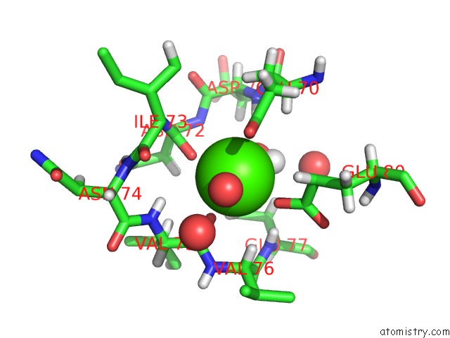
Mono view
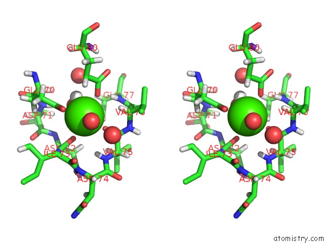
Stereo pair view
Mono view
Stereo pair view
A full contact list of Calcium with other atoms in the Ca binding
site number 1 of Cationic Trypsin in Its Apo Form (Deuterated Sample at 295 K) within 5.0Å range:
|
Reference:
J.Schiebel,
R.Gaspari,
T.Wulsdorf,
K.Ngo,
C.Sohn,
T.E.Schrader,
A.Cavalli,
A.Ostermann,
A.Heine,
G.Klebe.
Intriguing Role of Water in Protein-Ligand Binding Studied By Neutron Crystallography on Trypsin Complexes. Nat Commun V. 9 3559 2018.
ISSN: ESSN 2041-1723
PubMed: 30177695
DOI: 10.1038/S41467-018-05769-2
Page generated: Mon Jul 15 08:29:56 2024
ISSN: ESSN 2041-1723
PubMed: 30177695
DOI: 10.1038/S41467-018-05769-2
Last articles
Zn in 9MJ5Zn in 9HNW
Zn in 9G0L
Zn in 9FNE
Zn in 9DZN
Zn in 9E0I
Zn in 9D32
Zn in 9DAK
Zn in 8ZXC
Zn in 8ZUF