Calcium »
PDB 5mi5-5mop »
5mnx »
Calcium in PDB 5mnx: Neutron Structure of Cationic Trypsin in Complex with 2-Aminopyridine
Enzymatic activity of Neutron Structure of Cationic Trypsin in Complex with 2-Aminopyridine
All present enzymatic activity of Neutron Structure of Cationic Trypsin in Complex with 2-Aminopyridine:
3.4.21.4;
3.4.21.4;
Calcium Binding Sites:
The binding sites of Calcium atom in the Neutron Structure of Cationic Trypsin in Complex with 2-Aminopyridine
(pdb code 5mnx). This binding sites where shown within
5.0 Angstroms radius around Calcium atom.
In total only one binding site of Calcium was determined in the Neutron Structure of Cationic Trypsin in Complex with 2-Aminopyridine, PDB code: 5mnx:
In total only one binding site of Calcium was determined in the Neutron Structure of Cationic Trypsin in Complex with 2-Aminopyridine, PDB code: 5mnx:
Calcium binding site 1 out of 1 in 5mnx
Go back to
Calcium binding site 1 out
of 1 in the Neutron Structure of Cationic Trypsin in Complex with 2-Aminopyridine
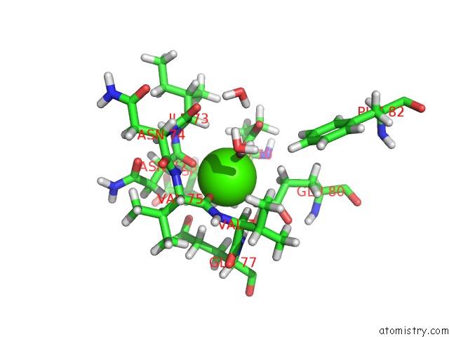
Mono view

Stereo pair view
Mono view
Stereo pair view
A full contact list of Calcium with other atoms in the Ca binding
site number 1 of Neutron Structure of Cationic Trypsin in Complex with 2-Aminopyridine within 5.0Å range:
|
Reference:
J.Schiebel,
R.Gaspari,
A.Sandner,
K.Ngo,
H.D.Gerber,
A.Cavalli,
A.Ostermann,
A.Heine,
G.Klebe.
Charges Shift Protonation: Neutron Diffraction Reveals That Aniline and 2-Aminopyridine Become Protonated Upon Binding to Trypsin. Angew. Chem. Int. Ed. Engl. V. 56 4887 2017.
ISSN: ESSN 1521-3773
PubMed: 28371253
DOI: 10.1002/ANIE.201701038
Page generated: Wed Jul 9 08:28:33 2025
ISSN: ESSN 1521-3773
PubMed: 28371253
DOI: 10.1002/ANIE.201701038
Last articles
F in 7M8VF in 7MCK
F in 7MAZ
F in 7MBO
F in 7MCE
F in 7MB2
F in 7MB1
F in 7MAX
F in 7M8R
F in 7M9R