Calcium »
PDB 5moq-5n2z »
5muv »
Calcium in PDB 5muv: Atomic Structure Fitted Into A Localized Reconstruction of Bacteriophage PHI6 Packaging Hexamer P4
Enzymatic activity of Atomic Structure Fitted Into A Localized Reconstruction of Bacteriophage PHI6 Packaging Hexamer P4
All present enzymatic activity of Atomic Structure Fitted Into A Localized Reconstruction of Bacteriophage PHI6 Packaging Hexamer P4:
3.6.1.15;
3.6.1.15;
Calcium Binding Sites:
The binding sites of Calcium atom in the Atomic Structure Fitted Into A Localized Reconstruction of Bacteriophage PHI6 Packaging Hexamer P4
(pdb code 5muv). This binding sites where shown within
5.0 Angstroms radius around Calcium atom.
In total 6 binding sites of Calcium where determined in the Atomic Structure Fitted Into A Localized Reconstruction of Bacteriophage PHI6 Packaging Hexamer P4, PDB code: 5muv:
Jump to Calcium binding site number: 1; 2; 3; 4; 5; 6;
In total 6 binding sites of Calcium where determined in the Atomic Structure Fitted Into A Localized Reconstruction of Bacteriophage PHI6 Packaging Hexamer P4, PDB code: 5muv:
Jump to Calcium binding site number: 1; 2; 3; 4; 5; 6;
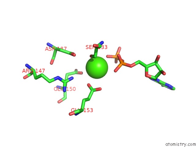
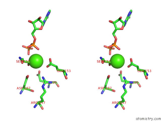
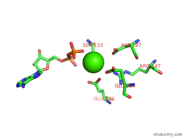
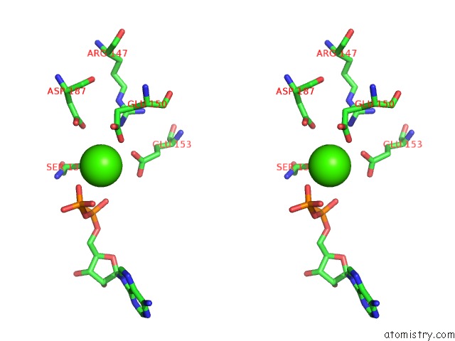
Calcium binding site 1 out of 6 in 5muv
Go back to
Calcium binding site 1 out
of 6 in the Atomic Structure Fitted Into A Localized Reconstruction of Bacteriophage PHI6 Packaging Hexamer P4
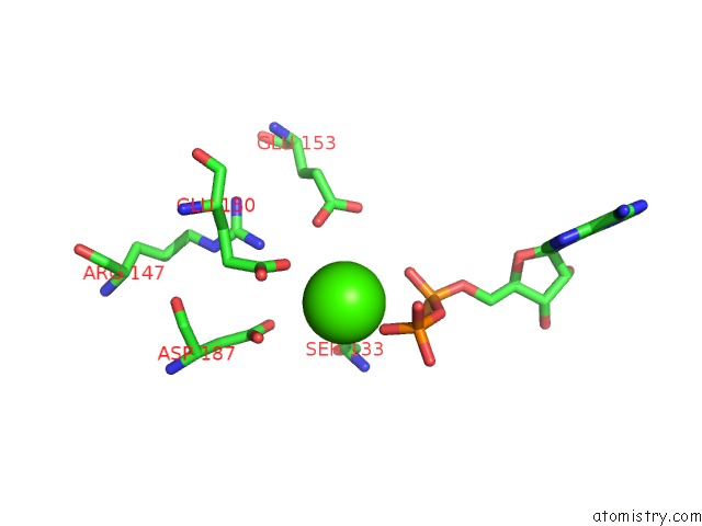
Mono view
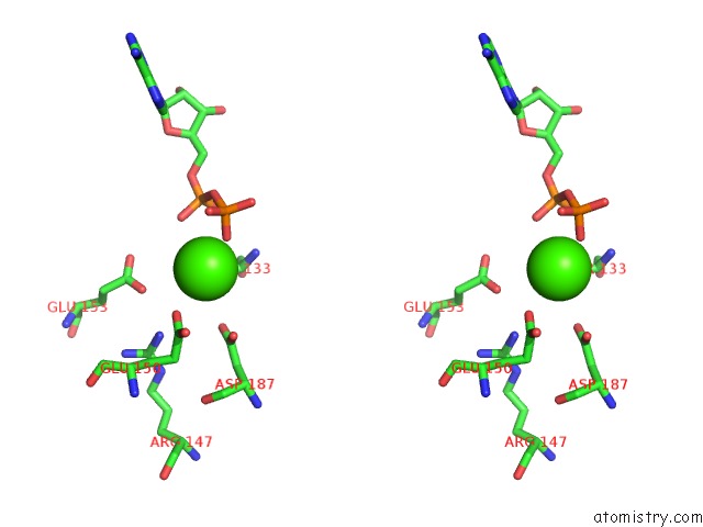
Stereo pair view
Mono view
Stereo pair view
A full contact list of Calcium with other atoms in the Ca binding
site number 1 of Atomic Structure Fitted Into A Localized Reconstruction of Bacteriophage PHI6 Packaging Hexamer P4 within 5.0Å range:
|
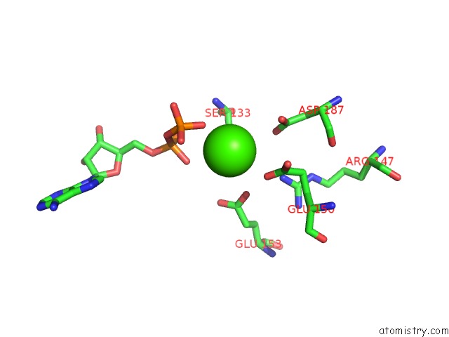
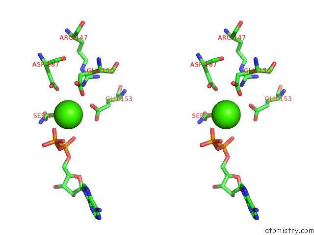
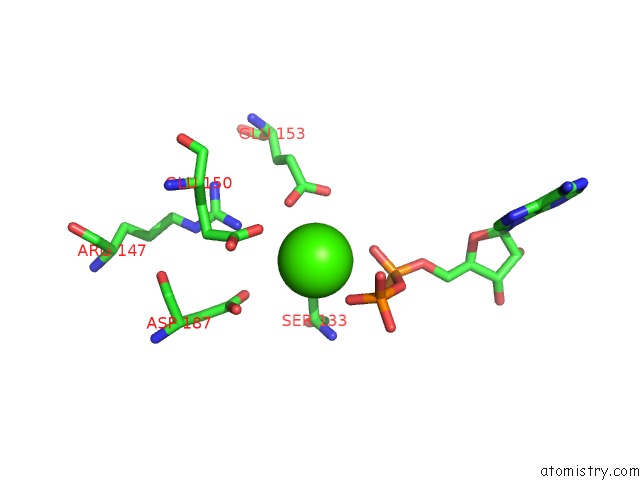
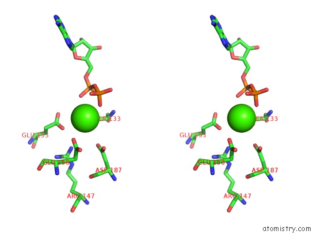
Calcium binding site 2 out of 6 in 5muv
Go back to
Calcium binding site 2 out
of 6 in the Atomic Structure Fitted Into A Localized Reconstruction of Bacteriophage PHI6 Packaging Hexamer P4
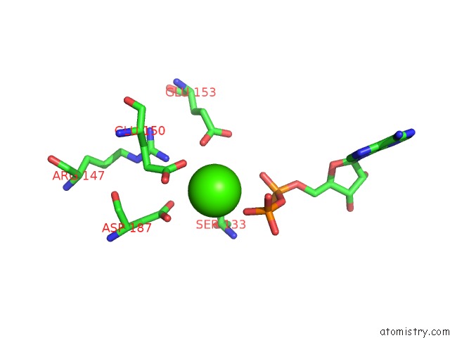
Mono view
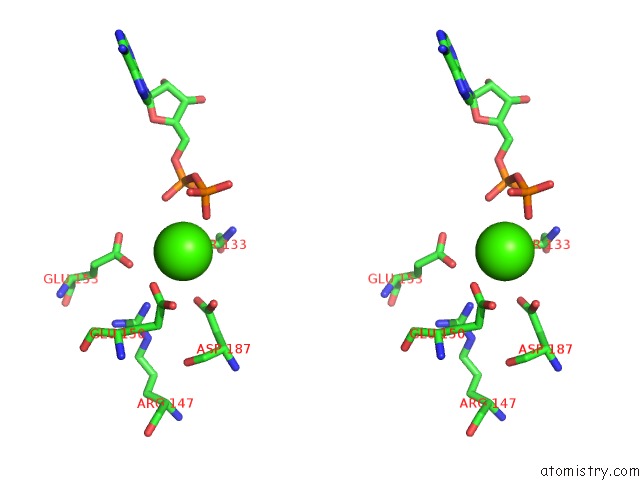
Stereo pair view
Mono view
Stereo pair view
A full contact list of Calcium with other atoms in the Ca binding
site number 2 of Atomic Structure Fitted Into A Localized Reconstruction of Bacteriophage PHI6 Packaging Hexamer P4 within 5.0Å range:
|
Calcium binding site 3 out of 6 in 5muv
Go back to
Calcium binding site 3 out
of 6 in the Atomic Structure Fitted Into A Localized Reconstruction of Bacteriophage PHI6 Packaging Hexamer P4
Mono view
Stereo pair view
Mono view
Stereo pair view
A full contact list of Calcium with other atoms in the Ca binding
site number 3 of Atomic Structure Fitted Into A Localized Reconstruction of Bacteriophage PHI6 Packaging Hexamer P4 within 5.0Å range:
|
Calcium binding site 4 out of 6 in 5muv
Go back to
Calcium binding site 4 out
of 6 in the Atomic Structure Fitted Into A Localized Reconstruction of Bacteriophage PHI6 Packaging Hexamer P4
Mono view
Stereo pair view
Mono view
Stereo pair view
A full contact list of Calcium with other atoms in the Ca binding
site number 4 of Atomic Structure Fitted Into A Localized Reconstruction of Bacteriophage PHI6 Packaging Hexamer P4 within 5.0Å range:
|
Calcium binding site 5 out of 6 in 5muv
Go back to
Calcium binding site 5 out
of 6 in the Atomic Structure Fitted Into A Localized Reconstruction of Bacteriophage PHI6 Packaging Hexamer P4
Mono view
Stereo pair view
Mono view
Stereo pair view
A full contact list of Calcium with other atoms in the Ca binding
site number 5 of Atomic Structure Fitted Into A Localized Reconstruction of Bacteriophage PHI6 Packaging Hexamer P4 within 5.0Å range:
|
Calcium binding site 6 out of 6 in 5muv
Go back to
Calcium binding site 6 out
of 6 in the Atomic Structure Fitted Into A Localized Reconstruction of Bacteriophage PHI6 Packaging Hexamer P4
Mono view
Stereo pair view
Mono view
Stereo pair view
A full contact list of Calcium with other atoms in the Ca binding
site number 6 of Atomic Structure Fitted Into A Localized Reconstruction of Bacteriophage PHI6 Packaging Hexamer P4 within 5.0Å range:
|
Reference:
Z.Sun,
K.El Omari,
X.Sun,
S.L.Ilca,
A.Kotecha,
D.I.Stuart,
M.M.Poranen,
J.T.Huiskonen.
Double-Stranded Rna Virus Outer Shell Assembly By Bona Fide Domain-Swapping. Nat Commun V. 8 14814 2017.
ISSN: ESSN 2041-1723
PubMed: 28287099
DOI: 10.1038/NCOMMS14814
Page generated: Mon Jul 15 08:37:54 2024
ISSN: ESSN 2041-1723
PubMed: 28287099
DOI: 10.1038/NCOMMS14814
Last articles
Zn in 9J0NZn in 9J0O
Zn in 9J0P
Zn in 9FJX
Zn in 9EKB
Zn in 9C0F
Zn in 9CAH
Zn in 9CH0
Zn in 9CH3
Zn in 9CH1