Calcium »
PDB 5npf-5odc »
5o2y »
Calcium in PDB 5o2y: uc(Nmr) Structure of the Calcium Bound Form of Pulg, Major Pseudopilin From Klebsiella Oxytoca T2SS
Calcium Binding Sites:
The binding sites of Calcium atom in the uc(Nmr) Structure of the Calcium Bound Form of Pulg, Major Pseudopilin From Klebsiella Oxytoca T2SS
(pdb code 5o2y). This binding sites where shown within
5.0 Angstroms radius around Calcium atom.
In total only one binding site of Calcium was determined in the uc(Nmr) Structure of the Calcium Bound Form of Pulg, Major Pseudopilin From Klebsiella Oxytoca T2SS, PDB code: 5o2y:
In total only one binding site of Calcium was determined in the uc(Nmr) Structure of the Calcium Bound Form of Pulg, Major Pseudopilin From Klebsiella Oxytoca T2SS, PDB code: 5o2y:
Calcium binding site 1 out of 1 in 5o2y
Go back to
Calcium binding site 1 out
of 1 in the uc(Nmr) Structure of the Calcium Bound Form of Pulg, Major Pseudopilin From Klebsiella Oxytoca T2SS
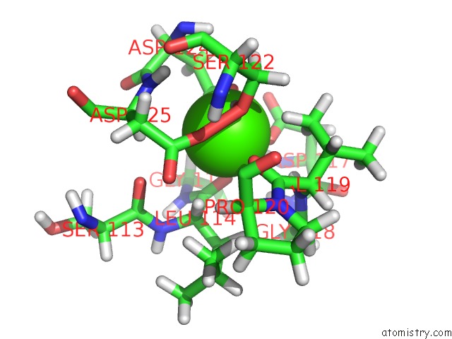
Mono view
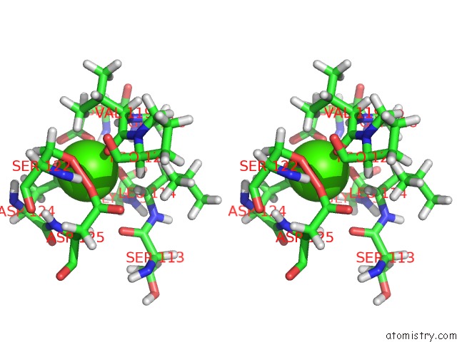
Stereo pair view
Mono view
Stereo pair view
A full contact list of Calcium with other atoms in the Ca binding
site number 1 of uc(Nmr) Structure of the Calcium Bound Form of Pulg, Major Pseudopilin From Klebsiella Oxytoca T2SS within 5.0Å range:
|
Reference:
A.Lopez-Castilla,
J.L.Thomassin,
B.Bardiaux,
W.Zheng,
M.Nivaskumar,
X.Yu,
M.Nilges,
E.H.Egelman,
N.Izadi-Pruneyre,
O.Francetic.
Structure of the Calcium-Dependent Type 2 Secretion Pseudopilus. Nat Microbiol V. 2 1686 2017.
ISSN: ESSN 2058-5276
PubMed: 28993624
DOI: 10.1038/S41564-017-0041-2
Page generated: Mon Jul 15 09:41:31 2024
ISSN: ESSN 2058-5276
PubMed: 28993624
DOI: 10.1038/S41564-017-0041-2
Last articles
Zn in 9J0NZn in 9J0O
Zn in 9J0P
Zn in 9FJX
Zn in 9EKB
Zn in 9C0F
Zn in 9CAH
Zn in 9CH0
Zn in 9CH3
Zn in 9CH1