Calcium »
PDB 5npf-5odc »
5o2z »
Calcium in PDB 5o2z: Domain Swap Dimer of the G167R Variant of Gelsolin Second Domain
Protein crystallography data
The structure of Domain Swap Dimer of the G167R Variant of Gelsolin Second Domain, PDB code: 5o2z
was solved by
F.Boni,
M.Milani,
E.Mastrangelo,
M.De Rosa,
with X-Ray Crystallography technique. A brief refinement statistics is given in the table below:
| Resolution Low / High (Å) | 45.39 / 1.70 |
| Space group | C 1 2 1 |
| Cell size a, b, c (Å), α, β, γ (°) | 106.290, 44.420, 58.010, 90.00, 110.13, 90.00 |
| R / Rfree (%) | 19.9 / 24.2 |
Other elements in 5o2z:
The structure of Domain Swap Dimer of the G167R Variant of Gelsolin Second Domain also contains other interesting chemical elements:
| Sodium | (Na) | 2 atoms |
Calcium Binding Sites:
The binding sites of Calcium atom in the Domain Swap Dimer of the G167R Variant of Gelsolin Second Domain
(pdb code 5o2z). This binding sites where shown within
5.0 Angstroms radius around Calcium atom.
In total 2 binding sites of Calcium where determined in the Domain Swap Dimer of the G167R Variant of Gelsolin Second Domain, PDB code: 5o2z:
Jump to Calcium binding site number: 1; 2;
In total 2 binding sites of Calcium where determined in the Domain Swap Dimer of the G167R Variant of Gelsolin Second Domain, PDB code: 5o2z:
Jump to Calcium binding site number: 1; 2;
Calcium binding site 1 out of 2 in 5o2z
Go back to
Calcium binding site 1 out
of 2 in the Domain Swap Dimer of the G167R Variant of Gelsolin Second Domain
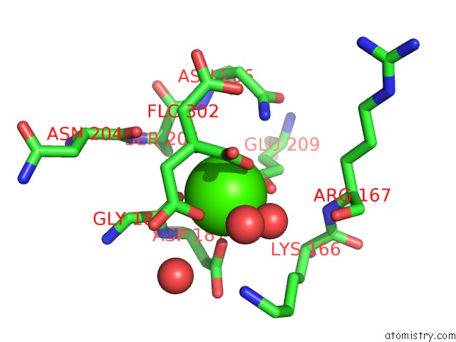
Mono view
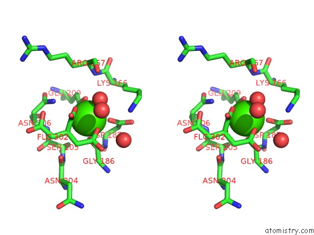
Stereo pair view
Mono view
Stereo pair view
A full contact list of Calcium with other atoms in the Ca binding
site number 1 of Domain Swap Dimer of the G167R Variant of Gelsolin Second Domain within 5.0Å range:
|
Calcium binding site 2 out of 2 in 5o2z
Go back to
Calcium binding site 2 out
of 2 in the Domain Swap Dimer of the G167R Variant of Gelsolin Second Domain
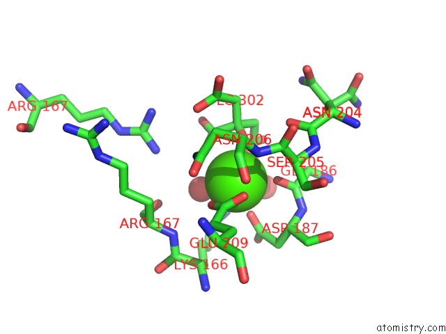
Mono view
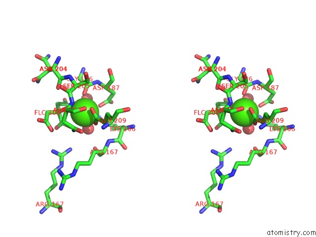
Stereo pair view
Mono view
Stereo pair view
A full contact list of Calcium with other atoms in the Ca binding
site number 2 of Domain Swap Dimer of the G167R Variant of Gelsolin Second Domain within 5.0Å range:
|
Reference:
F.Boni,
M.Milani,
A.Barbiroli,
L.Diomede,
E.Mastrangelo,
M.De Rosa.
Gelsolin Pathogenic GLY167ARG Mutation Promotes Domain-Swap Dimerization of the Protein. Hum. Mol. Genet. V. 27 53 2018.
ISSN: ESSN 1460-2083
PubMed: 29069428
DOI: 10.1093/HMG/DDX383
Page generated: Mon Jul 15 09:41:32 2024
ISSN: ESSN 1460-2083
PubMed: 29069428
DOI: 10.1093/HMG/DDX383
Last articles
Zn in 9J0NZn in 9J0O
Zn in 9J0P
Zn in 9FJX
Zn in 9EKB
Zn in 9C0F
Zn in 9CAH
Zn in 9CH0
Zn in 9CH3
Zn in 9CH1