Calcium »
PDB 5npf-5odc »
5o76 »
Calcium in PDB 5o76: Structure of PHOSPHOY371 C-Cbl in Complex with ZAP70-Peptide and Ubv.Pcbl Ubiquitin Variant
Enzymatic activity of Structure of PHOSPHOY371 C-Cbl in Complex with ZAP70-Peptide and Ubv.Pcbl Ubiquitin Variant
All present enzymatic activity of Structure of PHOSPHOY371 C-Cbl in Complex with ZAP70-Peptide and Ubv.Pcbl Ubiquitin Variant:
2.3.2.27;
2.3.2.27;
Protein crystallography data
The structure of Structure of PHOSPHOY371 C-Cbl in Complex with ZAP70-Peptide and Ubv.Pcbl Ubiquitin Variant, PDB code: 5o76
was solved by
M.Gabrielsen,
L.Buetow,
D.T.Huang,
with X-Ray Crystallography technique. A brief refinement statistics is given in the table below:
| Resolution Low / High (Å) | 35.34 / 2.47 |
| Space group | P 21 21 21 |
| Cell size a, b, c (Å), α, β, γ (°) | 94.790, 101.281, 117.339, 90.00, 90.00, 90.00 |
| R / Rfree (%) | 21.8 / 24.8 |
Other elements in 5o76:
The structure of Structure of PHOSPHOY371 C-Cbl in Complex with ZAP70-Peptide and Ubv.Pcbl Ubiquitin Variant also contains other interesting chemical elements:
| Zinc | (Zn) | 4 atoms |
Calcium Binding Sites:
The binding sites of Calcium atom in the Structure of PHOSPHOY371 C-Cbl in Complex with ZAP70-Peptide and Ubv.Pcbl Ubiquitin Variant
(pdb code 5o76). This binding sites where shown within
5.0 Angstroms radius around Calcium atom.
In total 2 binding sites of Calcium where determined in the Structure of PHOSPHOY371 C-Cbl in Complex with ZAP70-Peptide and Ubv.Pcbl Ubiquitin Variant, PDB code: 5o76:
Jump to Calcium binding site number: 1; 2;
In total 2 binding sites of Calcium where determined in the Structure of PHOSPHOY371 C-Cbl in Complex with ZAP70-Peptide and Ubv.Pcbl Ubiquitin Variant, PDB code: 5o76:
Jump to Calcium binding site number: 1; 2;
Calcium binding site 1 out of 2 in 5o76
Go back to
Calcium binding site 1 out
of 2 in the Structure of PHOSPHOY371 C-Cbl in Complex with ZAP70-Peptide and Ubv.Pcbl Ubiquitin Variant
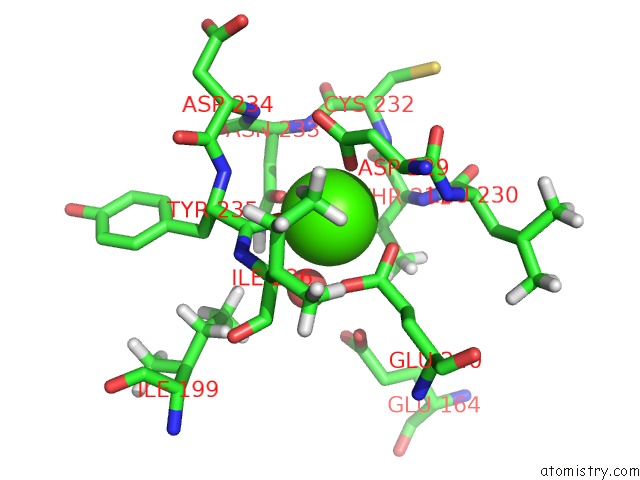
Mono view
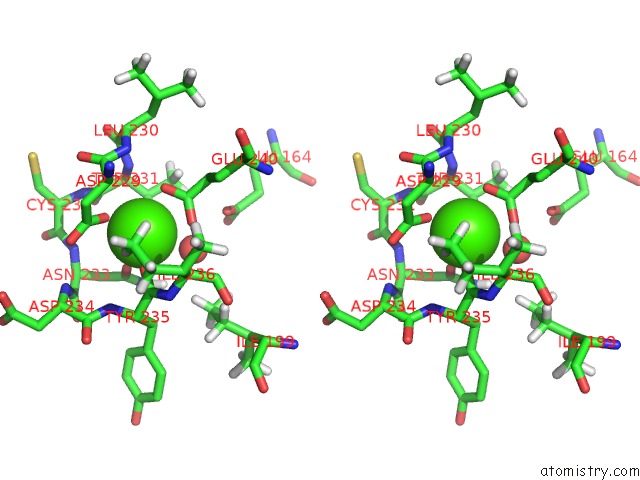
Stereo pair view
Mono view
Stereo pair view
A full contact list of Calcium with other atoms in the Ca binding
site number 1 of Structure of PHOSPHOY371 C-Cbl in Complex with ZAP70-Peptide and Ubv.Pcbl Ubiquitin Variant within 5.0Å range:
|
Calcium binding site 2 out of 2 in 5o76
Go back to
Calcium binding site 2 out
of 2 in the Structure of PHOSPHOY371 C-Cbl in Complex with ZAP70-Peptide and Ubv.Pcbl Ubiquitin Variant
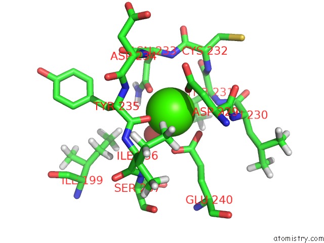
Mono view
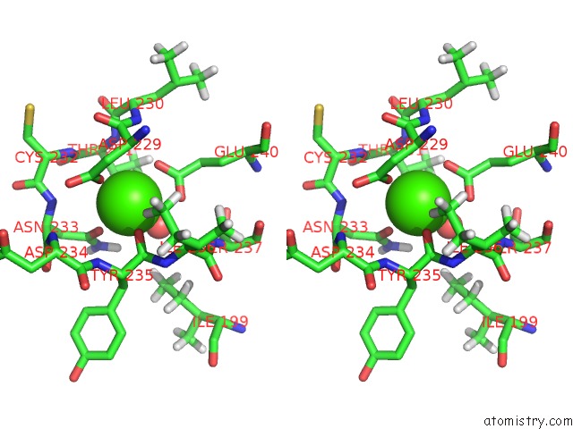
Stereo pair view
Mono view
Stereo pair view
A full contact list of Calcium with other atoms in the Ca binding
site number 2 of Structure of PHOSPHOY371 C-Cbl in Complex with ZAP70-Peptide and Ubv.Pcbl Ubiquitin Variant within 5.0Å range:
|
Reference:
M.Gabrielsen,
L.Buetow,
M.A.Nakasone,
S.F.Ahmed,
G.J.Sibbet,
B.O.Smith,
W.Zhang,
S.S.Sidhu,
D.T.Huang.
A General Strategy For Discovery of Inhibitors and Activators of Ring and U-Box E3 Ligases with Ubiquitin Variants. Mol. Cell V. 68 456 2017.
ISSN: ISSN 1097-4164
PubMed: 29053960
DOI: 10.1016/J.MOLCEL.2017.09.027
Page generated: Wed Jul 9 09:14:45 2025
ISSN: ISSN 1097-4164
PubMed: 29053960
DOI: 10.1016/J.MOLCEL.2017.09.027
Last articles
Fe in 2YXOFe in 2YRS
Fe in 2YXC
Fe in 2YNM
Fe in 2YVJ
Fe in 2YP1
Fe in 2YU2
Fe in 2YU1
Fe in 2YQB
Fe in 2YOO