Calcium »
PDB 5npf-5odc »
5o8x »
Calcium in PDB 5o8x: The X-Ray Structure of Catenated Lytic Transglycosylase SLTB1 From Pseudomonas Aeruginosa
Protein crystallography data
The structure of The X-Ray Structure of Catenated Lytic Transglycosylase SLTB1 From Pseudomonas Aeruginosa, PDB code: 5o8x
was solved by
T.Dominguez-Gil,
R.Molina,
with X-Ray Crystallography technique. A brief refinement statistics is given in the table below:
| Resolution Low / High (Å) | 48.26 / 2.50 |
| Space group | C 1 2 1 |
| Cell size a, b, c (Å), α, β, γ (°) | 115.064, 116.358, 54.865, 90.00, 118.41, 90.00 |
| R / Rfree (%) | 17.7 / 22.9 |
Other elements in 5o8x:
The structure of The X-Ray Structure of Catenated Lytic Transglycosylase SLTB1 From Pseudomonas Aeruginosa also contains other interesting chemical elements:
| Arsenic | (As) | 2 atoms |
Calcium Binding Sites:
The binding sites of Calcium atom in the The X-Ray Structure of Catenated Lytic Transglycosylase SLTB1 From Pseudomonas Aeruginosa
(pdb code 5o8x). This binding sites where shown within
5.0 Angstroms radius around Calcium atom.
In total 2 binding sites of Calcium where determined in the The X-Ray Structure of Catenated Lytic Transglycosylase SLTB1 From Pseudomonas Aeruginosa, PDB code: 5o8x:
Jump to Calcium binding site number: 1; 2;
In total 2 binding sites of Calcium where determined in the The X-Ray Structure of Catenated Lytic Transglycosylase SLTB1 From Pseudomonas Aeruginosa, PDB code: 5o8x:
Jump to Calcium binding site number: 1; 2;
Calcium binding site 1 out of 2 in 5o8x
Go back to
Calcium binding site 1 out
of 2 in the The X-Ray Structure of Catenated Lytic Transglycosylase SLTB1 From Pseudomonas Aeruginosa
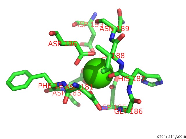
Mono view
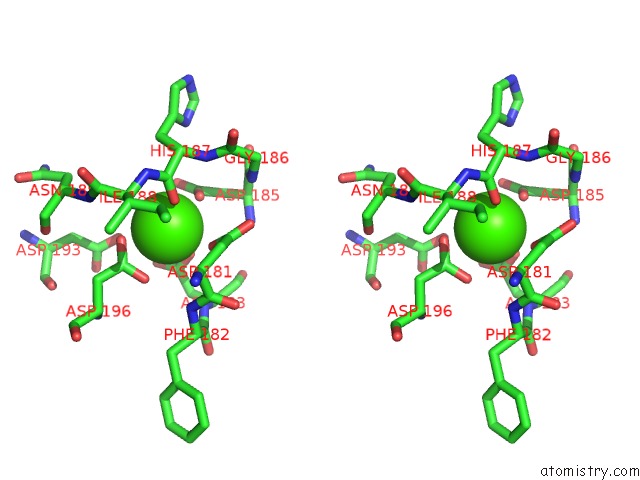
Stereo pair view
Mono view
Stereo pair view
A full contact list of Calcium with other atoms in the Ca binding
site number 1 of The X-Ray Structure of Catenated Lytic Transglycosylase SLTB1 From Pseudomonas Aeruginosa within 5.0Å range:
|
Calcium binding site 2 out of 2 in 5o8x
Go back to
Calcium binding site 2 out
of 2 in the The X-Ray Structure of Catenated Lytic Transglycosylase SLTB1 From Pseudomonas Aeruginosa
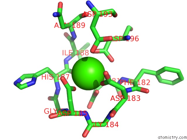
Mono view
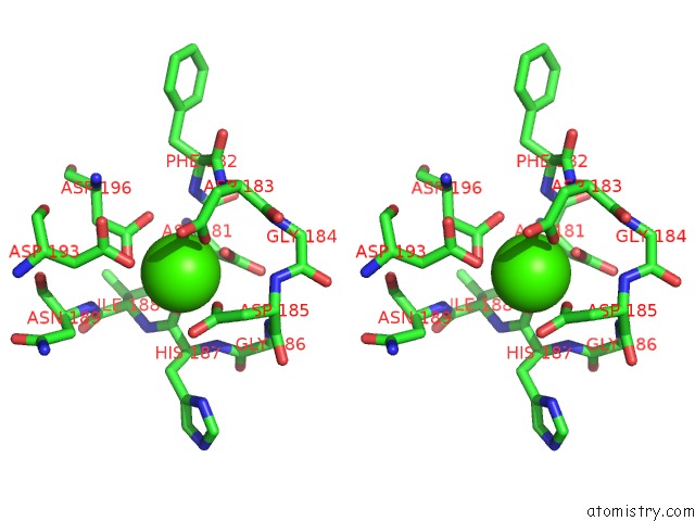
Stereo pair view
Mono view
Stereo pair view
A full contact list of Calcium with other atoms in the Ca binding
site number 2 of The X-Ray Structure of Catenated Lytic Transglycosylase SLTB1 From Pseudomonas Aeruginosa within 5.0Å range:
|
Reference:
T.Dominguez-Gil,
R.Molina,
D.A.Dik,
E.Spink,
S.Mobashery,
J.A.Hermoso.
X-Ray Structure of Catenated Lytic Transglycosylase SLTB1. Biochemistry V. 56 6317 2017.
ISSN: ISSN 1520-4995
PubMed: 29131935
DOI: 10.1021/ACS.BIOCHEM.7B00932
Page generated: Mon Jul 15 09:44:06 2024
ISSN: ISSN 1520-4995
PubMed: 29131935
DOI: 10.1021/ACS.BIOCHEM.7B00932
Last articles
Zn in 9J0NZn in 9J0O
Zn in 9J0P
Zn in 9FJX
Zn in 9EKB
Zn in 9C0F
Zn in 9CAH
Zn in 9CH0
Zn in 9CH3
Zn in 9CH1