Calcium »
PDB 5odu-5oyl »
5ov7 »
Calcium in PDB 5ov7: Tubulin - Rigosertib Complex
Protein crystallography data
The structure of Tubulin - Rigosertib Complex, PDB code: 5ov7
was solved by
G.Menchon,
A.E.Prota,
M.Steinmetz,
M.Jost,
with X-Ray Crystallography technique. A brief refinement statistics is given in the table below:
| Resolution Low / High (Å) | 48.08 / 2.40 |
| Space group | P 21 21 21 |
| Cell size a, b, c (Å), α, β, γ (°) | 104.732, 156.765, 182.629, 90.00, 90.00, 90.00 |
| R / Rfree (%) | 19 / 23.5 |
Other elements in 5ov7:
The structure of Tubulin - Rigosertib Complex also contains other interesting chemical elements:
| Magnesium | (Mg) | 5 atoms |
Calcium Binding Sites:
The binding sites of Calcium atom in the Tubulin - Rigosertib Complex
(pdb code 5ov7). This binding sites where shown within
5.0 Angstroms radius around Calcium atom.
In total only one binding site of Calcium was determined in the Tubulin - Rigosertib Complex, PDB code: 5ov7:
In total only one binding site of Calcium was determined in the Tubulin - Rigosertib Complex, PDB code: 5ov7:
Calcium binding site 1 out of 1 in 5ov7
Go back to
Calcium binding site 1 out
of 1 in the Tubulin - Rigosertib Complex
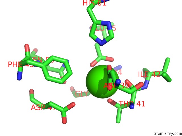
Mono view
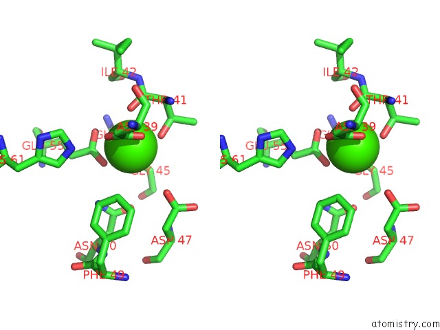
Stereo pair view
Mono view
Stereo pair view
A full contact list of Calcium with other atoms in the Ca binding
site number 1 of Tubulin - Rigosertib Complex within 5.0Å range:
|
Reference:
M.Jost,
Y.Chen,
L.A.Gilbert,
M.A.Horlbeck,
L.Krenning,
G.Menchon,
A.Rai,
M.Y.Cho,
J.J.Stern,
A.E.Prota,
M.Kampmann,
A.Akhmanova,
M.O.Steinmetz,
M.E.Tanenbaum,
J.S.Weissman.
Combined Crispri/A-Based Chemical Genetic Screens Reveal That Rigosertib Is A Microtubule-Destabilizing Agent. Mol. Cell V. 68 210 2017.
ISSN: ISSN 1097-4164
PubMed: 28985505
DOI: 10.1016/J.MOLCEL.2017.09.012
Page generated: Mon Jul 15 09:53:39 2024
ISSN: ISSN 1097-4164
PubMed: 28985505
DOI: 10.1016/J.MOLCEL.2017.09.012
Last articles
Zn in 9J0NZn in 9J0O
Zn in 9J0P
Zn in 9FJX
Zn in 9EKB
Zn in 9C0F
Zn in 9CAH
Zn in 9CH0
Zn in 9CH3
Zn in 9CH1