Calcium »
PDB 5odu-5oyl »
5oy0 »
Calcium in PDB 5oy0: Structure of Synechocystis Photosystem I Trimer at 2.5A Resolution
Enzymatic activity of Structure of Synechocystis Photosystem I Trimer at 2.5A Resolution
All present enzymatic activity of Structure of Synechocystis Photosystem I Trimer at 2.5A Resolution:
1.97.1.12;
1.97.1.12;
Protein crystallography data
The structure of Structure of Synechocystis Photosystem I Trimer at 2.5A Resolution, PDB code: 5oy0
was solved by
N.Nelson,
T.Malavath,
I.Caspy,
with X-Ray Crystallography technique. A brief refinement statistics is given in the table below:
| Resolution Low / High (Å) | 49.48 / 2.50 |
| Space group | P 1 21 1 |
| Cell size a, b, c (Å), α, β, γ (°) | 212.170, 137.622, 225.095, 90.00, 116.74, 90.00 |
| R / Rfree (%) | 22.8 / 26.4 |
Other elements in 5oy0:
The structure of Structure of Synechocystis Photosystem I Trimer at 2.5A Resolution also contains other interesting chemical elements:
| Magnesium | (Mg) | 287 atoms |
| Iron | (Fe) | 36 atoms |
| Chlorine | (Cl) | 3 atoms |
Calcium Binding Sites:
The binding sites of Calcium atom in the Structure of Synechocystis Photosystem I Trimer at 2.5A Resolution
(pdb code 5oy0). This binding sites where shown within
5.0 Angstroms radius around Calcium atom.
In total 6 binding sites of Calcium where determined in the Structure of Synechocystis Photosystem I Trimer at 2.5A Resolution, PDB code: 5oy0:
Jump to Calcium binding site number: 1; 2; 3; 4; 5; 6;
In total 6 binding sites of Calcium where determined in the Structure of Synechocystis Photosystem I Trimer at 2.5A Resolution, PDB code: 5oy0:
Jump to Calcium binding site number: 1; 2; 3; 4; 5; 6;
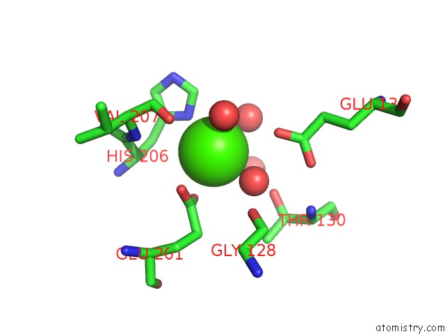
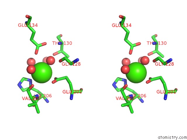
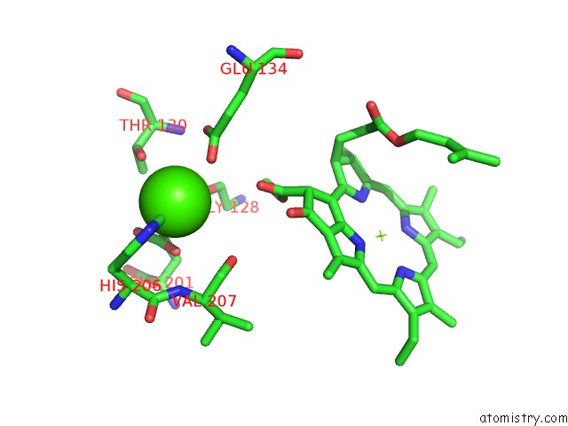
Calcium binding site 1 out of 6 in 5oy0
Go back to
Calcium binding site 1 out
of 6 in the Structure of Synechocystis Photosystem I Trimer at 2.5A Resolution
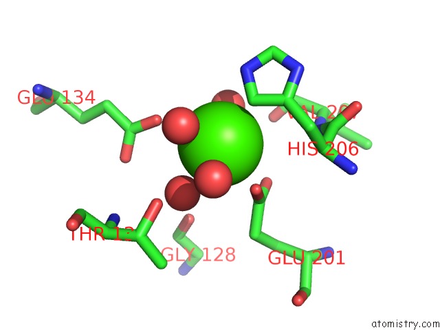
Mono view
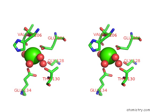
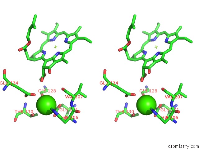
Stereo pair view
Mono view
Stereo pair view
A full contact list of Calcium with other atoms in the Ca binding
site number 1 of Structure of Synechocystis Photosystem I Trimer at 2.5A Resolution within 5.0Å range:
|
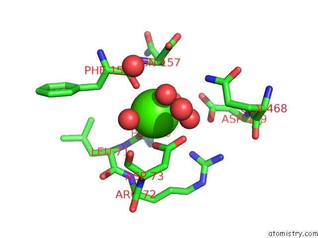
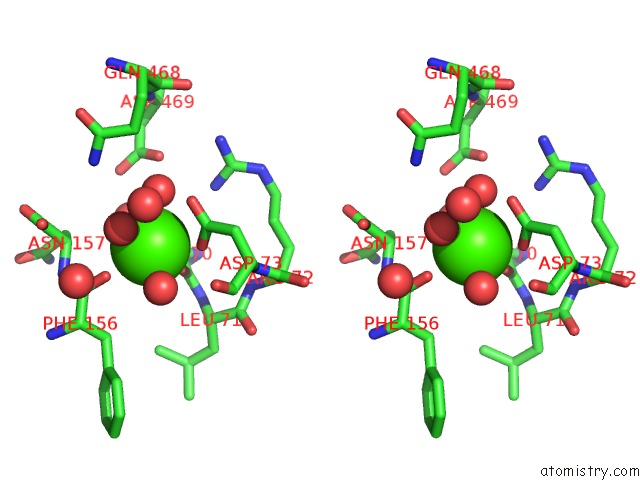
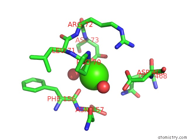
Calcium binding site 2 out of 6 in 5oy0
Go back to
Calcium binding site 2 out
of 6 in the Structure of Synechocystis Photosystem I Trimer at 2.5A Resolution
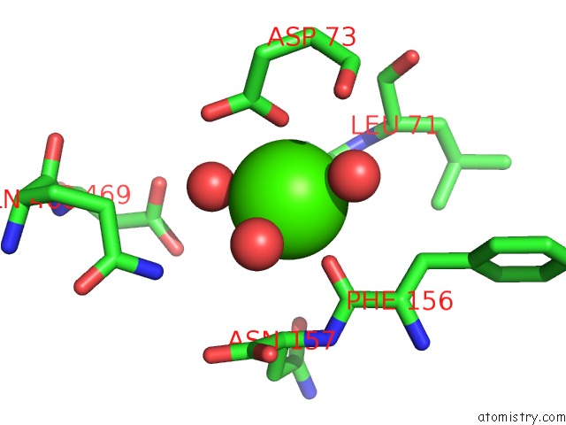
Mono view
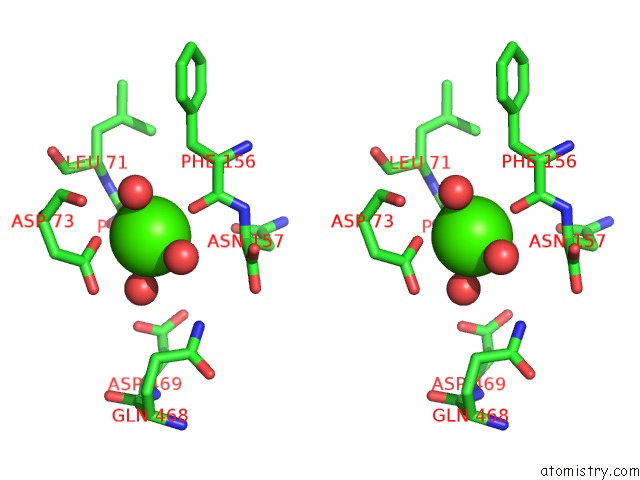
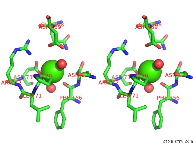
Stereo pair view
Mono view
Stereo pair view
A full contact list of Calcium with other atoms in the Ca binding
site number 2 of Structure of Synechocystis Photosystem I Trimer at 2.5A Resolution within 5.0Å range:
|
Calcium binding site 3 out of 6 in 5oy0
Go back to
Calcium binding site 3 out
of 6 in the Structure of Synechocystis Photosystem I Trimer at 2.5A Resolution
Mono view
Stereo pair view
Mono view
Stereo pair view
A full contact list of Calcium with other atoms in the Ca binding
site number 3 of Structure of Synechocystis Photosystem I Trimer at 2.5A Resolution within 5.0Å range:
|
Calcium binding site 4 out of 6 in 5oy0
Go back to
Calcium binding site 4 out
of 6 in the Structure of Synechocystis Photosystem I Trimer at 2.5A Resolution
Mono view
Stereo pair view
Mono view
Stereo pair view
A full contact list of Calcium with other atoms in the Ca binding
site number 4 of Structure of Synechocystis Photosystem I Trimer at 2.5A Resolution within 5.0Å range:
|
Calcium binding site 5 out of 6 in 5oy0
Go back to
Calcium binding site 5 out
of 6 in the Structure of Synechocystis Photosystem I Trimer at 2.5A Resolution
Mono view
Stereo pair view
Mono view
Stereo pair view
A full contact list of Calcium with other atoms in the Ca binding
site number 5 of Structure of Synechocystis Photosystem I Trimer at 2.5A Resolution within 5.0Å range:
|
Calcium binding site 6 out of 6 in 5oy0
Go back to
Calcium binding site 6 out
of 6 in the Structure of Synechocystis Photosystem I Trimer at 2.5A Resolution
Mono view
Stereo pair view
Mono view
Stereo pair view
A full contact list of Calcium with other atoms in the Ca binding
site number 6 of Structure of Synechocystis Photosystem I Trimer at 2.5A Resolution within 5.0Å range:
|
Reference:
T.Malavath,
I.Caspy,
S.Y.Netzer-El,
D.Klaiman,
N.Nelson.
Structure and Function of Wild-Type and Subunit-Depleted Photosystem I in Synechocystis. Biochim. Biophys. Acta V.1859 645 2018.
ISSN: ISSN 0006-3002
PubMed: 29414678
DOI: 10.1016/J.BBABIO.2018.02.002
Page generated: Wed Jul 9 09:23:02 2025
ISSN: ISSN 0006-3002
PubMed: 29414678
DOI: 10.1016/J.BBABIO.2018.02.002
Last articles
Cl in 5I4UCl in 5I4K
Cl in 5I4Q
Cl in 5I1T
Cl in 5I3W
Cl in 5I21
Cl in 5HZ9
Cl in 5I1F
Cl in 5I0D
Cl in 5I13