Calcium »
PDB 5tae-5tua »
5ti8 »
Calcium in PDB 5ti8: Crystal Structure of An Aspartate Aminotransferase From Pseudomonas
Protein crystallography data
The structure of Crystal Structure of An Aspartate Aminotransferase From Pseudomonas, PDB code: 5ti8
was solved by
T.S.Peat,
J.Newman,
with X-Ray Crystallography technique. A brief refinement statistics is given in the table below:
| Resolution Low / High (Å) | 47.70 / 2.07 |
| Space group | I 1 2 1 |
| Cell size a, b, c (Å), α, β, γ (°) | 76.444, 95.429, 114.449, 90.00, 100.21, 90.00 |
| R / Rfree (%) | 18.4 / 23.8 |
Calcium Binding Sites:
The binding sites of Calcium atom in the Crystal Structure of An Aspartate Aminotransferase From Pseudomonas
(pdb code 5ti8). This binding sites where shown within
5.0 Angstroms radius around Calcium atom.
In total 2 binding sites of Calcium where determined in the Crystal Structure of An Aspartate Aminotransferase From Pseudomonas, PDB code: 5ti8:
Jump to Calcium binding site number: 1; 2;
In total 2 binding sites of Calcium where determined in the Crystal Structure of An Aspartate Aminotransferase From Pseudomonas, PDB code: 5ti8:
Jump to Calcium binding site number: 1; 2;
Calcium binding site 1 out of 2 in 5ti8
Go back to
Calcium binding site 1 out
of 2 in the Crystal Structure of An Aspartate Aminotransferase From Pseudomonas
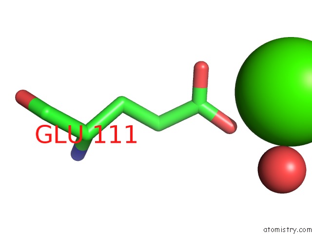
Mono view
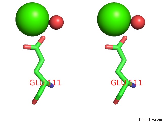
Stereo pair view
Mono view
Stereo pair view
A full contact list of Calcium with other atoms in the Ca binding
site number 1 of Crystal Structure of An Aspartate Aminotransferase From Pseudomonas within 5.0Å range:
|
Calcium binding site 2 out of 2 in 5ti8
Go back to
Calcium binding site 2 out
of 2 in the Crystal Structure of An Aspartate Aminotransferase From Pseudomonas
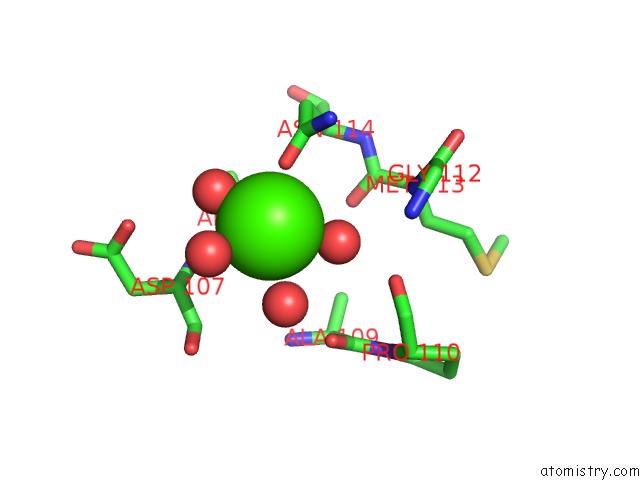
Mono view
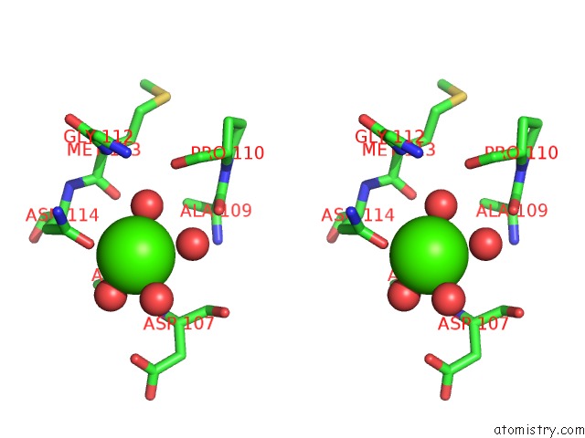
Stereo pair view
Mono view
Stereo pair view
A full contact list of Calcium with other atoms in the Ca binding
site number 2 of Crystal Structure of An Aspartate Aminotransferase From Pseudomonas within 5.0Å range:
|
Reference:
M.Wilding,
C.Scott,
J.Newman,
T.S.Peat.
Crystal Structure of A Putrescine Aminotransferase From Pseudomonas Sp. Strain Aac. Acta Crystallogr F Struct V. 73 29 2017BIOL Commun.
ISSN: ESSN 2053-230X
PubMed: 28045391
DOI: 10.1107/S2053230X16019658
Page generated: Mon Jul 15 11:18:20 2024
ISSN: ESSN 2053-230X
PubMed: 28045391
DOI: 10.1107/S2053230X16019658
Last articles
Zn in 9J0NZn in 9J0O
Zn in 9J0P
Zn in 9FJX
Zn in 9EKB
Zn in 9C0F
Zn in 9CAH
Zn in 9CH0
Zn in 9CH3
Zn in 9CH1