Calcium »
PDB 5tad-5ttc »
5tr2 »
Calcium in PDB 5tr2: Crystal Structure of the D263G Missense Variant of Human PGM1
Enzymatic activity of Crystal Structure of the D263G Missense Variant of Human PGM1
All present enzymatic activity of Crystal Structure of the D263G Missense Variant of Human PGM1:
5.4.2.2;
5.4.2.2;
Protein crystallography data
The structure of Crystal Structure of the D263G Missense Variant of Human PGM1, PDB code: 5tr2
was solved by
L.J.Beamer,
K.M.Stiers,
with X-Ray Crystallography technique. A brief refinement statistics is given in the table below:
| Resolution Low / High (Å) | 51.84 / 2.50 |
| Space group | P 41 21 2 |
| Cell size a, b, c (Å), α, β, γ (°) | 172.124, 172.124, 99.005, 90.00, 90.00, 90.00 |
| R / Rfree (%) | 22 / 29.2 |
Calcium Binding Sites:
The binding sites of Calcium atom in the Crystal Structure of the D263G Missense Variant of Human PGM1
(pdb code 5tr2). This binding sites where shown within
5.0 Angstroms radius around Calcium atom.
In total 2 binding sites of Calcium where determined in the Crystal Structure of the D263G Missense Variant of Human PGM1, PDB code: 5tr2:
Jump to Calcium binding site number: 1; 2;
In total 2 binding sites of Calcium where determined in the Crystal Structure of the D263G Missense Variant of Human PGM1, PDB code: 5tr2:
Jump to Calcium binding site number: 1; 2;
Calcium binding site 1 out of 2 in 5tr2
Go back to
Calcium binding site 1 out
of 2 in the Crystal Structure of the D263G Missense Variant of Human PGM1
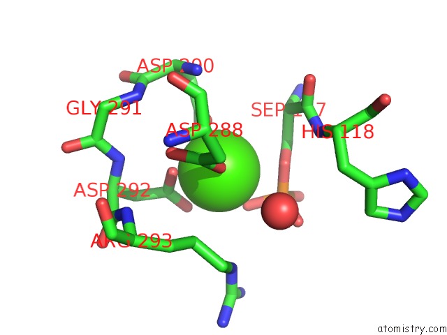
Mono view
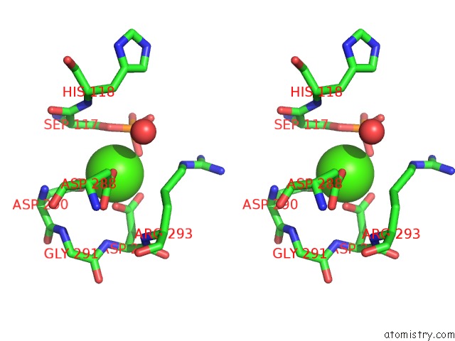
Stereo pair view
Mono view
Stereo pair view
A full contact list of Calcium with other atoms in the Ca binding
site number 1 of Crystal Structure of the D263G Missense Variant of Human PGM1 within 5.0Å range:
|
Calcium binding site 2 out of 2 in 5tr2
Go back to
Calcium binding site 2 out
of 2 in the Crystal Structure of the D263G Missense Variant of Human PGM1
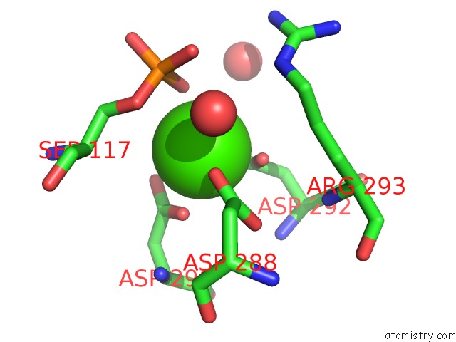
Mono view
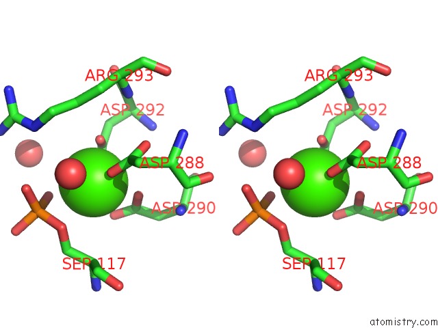
Stereo pair view
Mono view
Stereo pair view
A full contact list of Calcium with other atoms in the Ca binding
site number 2 of Crystal Structure of the D263G Missense Variant of Human PGM1 within 5.0Å range:
|
Reference:
K.M.Stiers,
A.C.Graham,
B.N.Kain,
L.J.Beamer.
ASP263 Missense Variants Perturb the Active Site of Human Phosphoglucomutase 1. Febs J. V. 284 937 2017.
ISSN: ISSN 1742-4658
PubMed: 28117557
DOI: 10.1111/FEBS.14025
Page generated: Mon Jul 15 11:23:03 2024
ISSN: ISSN 1742-4658
PubMed: 28117557
DOI: 10.1111/FEBS.14025
Last articles
Zn in 9JYWZn in 9IR4
Zn in 9IR3
Zn in 9GMX
Zn in 9GMW
Zn in 9JEJ
Zn in 9ERF
Zn in 9ERE
Zn in 9EGV
Zn in 9EGW