Calcium »
PDB 5tvq-5ufe »
5tx6 »
Calcium in PDB 5tx6: Structure of Tgf-BETA2 Derivative with Deletion of Residues 52-71 and 10 Single Amino Acid Mutations (Mmtgf-BETA2-7M)
Protein crystallography data
The structure of Structure of Tgf-BETA2 Derivative with Deletion of Residues 52-71 and 10 Single Amino Acid Mutations (Mmtgf-BETA2-7M), PDB code: 5tx6
was solved by
E.M.Petrunak,
A.P.Hinck,
with X-Ray Crystallography technique. A brief refinement statistics is given in the table below:
| Resolution Low / High (Å) | 36.48 / 2.75 |
| Space group | P 31 2 1 |
| Cell size a, b, c (Å), α, β, γ (°) | 81.744, 81.744, 80.926, 90.00, 90.00, 120.00 |
| R / Rfree (%) | 21.3 / 27.2 |
Calcium Binding Sites:
The binding sites of Calcium atom in the Structure of Tgf-BETA2 Derivative with Deletion of Residues 52-71 and 10 Single Amino Acid Mutations (Mmtgf-BETA2-7M)
(pdb code 5tx6). This binding sites where shown within
5.0 Angstroms radius around Calcium atom.
In total only one binding site of Calcium was determined in the Structure of Tgf-BETA2 Derivative with Deletion of Residues 52-71 and 10 Single Amino Acid Mutations (Mmtgf-BETA2-7M), PDB code: 5tx6:
In total only one binding site of Calcium was determined in the Structure of Tgf-BETA2 Derivative with Deletion of Residues 52-71 and 10 Single Amino Acid Mutations (Mmtgf-BETA2-7M), PDB code: 5tx6:
Calcium binding site 1 out of 1 in 5tx6
Go back to
Calcium binding site 1 out
of 1 in the Structure of Tgf-BETA2 Derivative with Deletion of Residues 52-71 and 10 Single Amino Acid Mutations (Mmtgf-BETA2-7M)
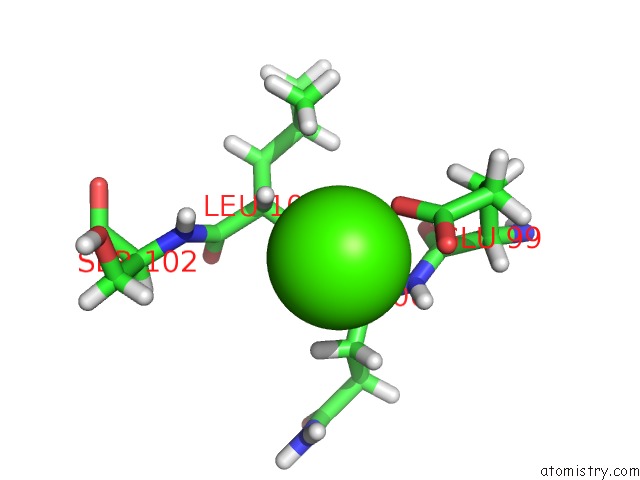
Mono view
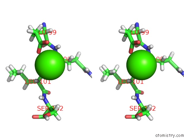
Stereo pair view
Mono view
Stereo pair view
A full contact list of Calcium with other atoms in the Ca binding
site number 1 of Structure of Tgf-BETA2 Derivative with Deletion of Residues 52-71 and 10 Single Amino Acid Mutations (Mmtgf-BETA2-7M) within 5.0Å range:
|
Reference:
S.K.Kim,
L.Barron,
C.S.Hinck,
E.M.Petrunak,
K.E.Cano,
A.Thangirala,
B.Iskra,
M.Brothers,
M.Vonberg,
B.Leal,
B.Richter,
R.Kodali,
A.B.Taylor,
S.Du,
C.O.Barnes,
T.Sulea,
G.Calero,
P.J.Hart,
M.J.Hart,
B.Demeler,
A.P.Hinck.
An Engineered Transforming Growth Factor Beta (Tgf-Beta ) Monomer That Functions As A Dominant Negative to Block Tgf-Beta Signaling. J. Biol. Chem. V. 292 7173 2017.
ISSN: ESSN 1083-351X
PubMed: 28228478
DOI: 10.1074/JBC.M116.768754
Page generated: Mon Jul 15 11:25:01 2024
ISSN: ESSN 1083-351X
PubMed: 28228478
DOI: 10.1074/JBC.M116.768754
Last articles
Zn in 9MJ5Zn in 9HNW
Zn in 9G0L
Zn in 9FNE
Zn in 9DZN
Zn in 9E0I
Zn in 9D32
Zn in 9DAK
Zn in 8ZXC
Zn in 8ZUF