Calcium »
PDB 5ufq-5uw6 »
5ug4 »
Calcium in PDB 5ug4: Structure of Spermidine N-Acetyltransferase Speg From Vibrio Cholerae
Enzymatic activity of Structure of Spermidine N-Acetyltransferase Speg From Vibrio Cholerae
All present enzymatic activity of Structure of Spermidine N-Acetyltransferase Speg From Vibrio Cholerae:
2.3.1.57;
2.3.1.57;
Protein crystallography data
The structure of Structure of Spermidine N-Acetyltransferase Speg From Vibrio Cholerae, PDB code: 5ug4
was solved by
E.V.Filippova,
G.Minasov,
L.Shuvalova,
O.Kiryukhina,
W.F.Anderson,
Centerfor Structural Genomics Of Infectious Diseases (Csgid),
with X-Ray Crystallography technique. A brief refinement statistics is given in the table below:
| Resolution Low / High (Å) | 30.00 / 2.15 |
| Space group | I 2 2 2 |
| Cell size a, b, c (Å), α, β, γ (°) | 71.884, 134.635, 137.346, 90.00, 90.00, 90.00 |
| R / Rfree (%) | 16.9 / 20 |
Calcium Binding Sites:
The binding sites of Calcium atom in the Structure of Spermidine N-Acetyltransferase Speg From Vibrio Cholerae
(pdb code 5ug4). This binding sites where shown within
5.0 Angstroms radius around Calcium atom.
In total 9 binding sites of Calcium where determined in the Structure of Spermidine N-Acetyltransferase Speg From Vibrio Cholerae, PDB code: 5ug4:
Jump to Calcium binding site number: 1; 2; 3; 4; 5; 6; 7; 8; 9;
In total 9 binding sites of Calcium where determined in the Structure of Spermidine N-Acetyltransferase Speg From Vibrio Cholerae, PDB code: 5ug4:
Jump to Calcium binding site number: 1; 2; 3; 4; 5; 6; 7; 8; 9;
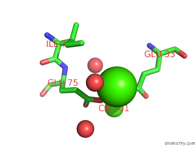
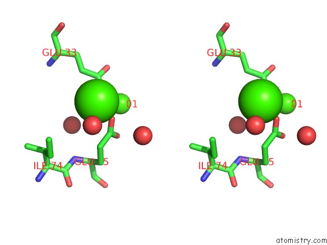
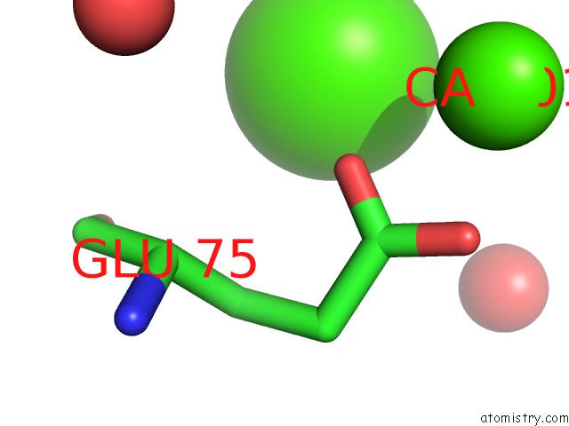
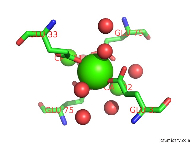
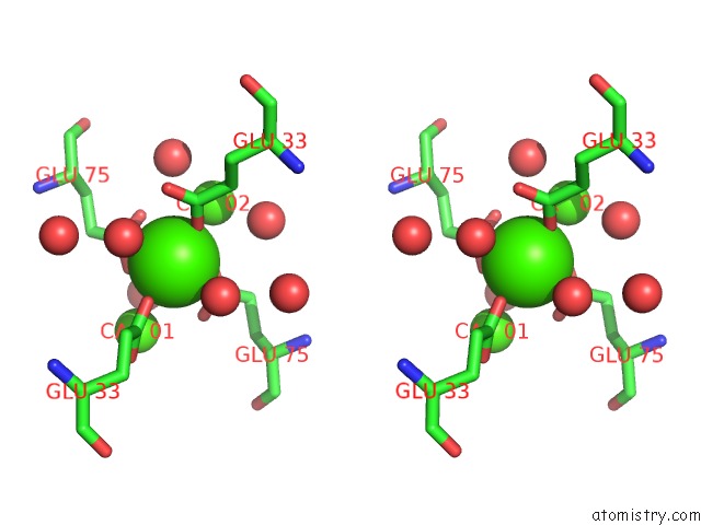
Calcium binding site 1 out of 9 in 5ug4
Go back to
Calcium binding site 1 out
of 9 in the Structure of Spermidine N-Acetyltransferase Speg From Vibrio Cholerae

Mono view
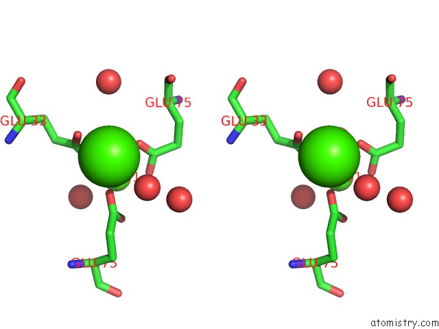
Stereo pair view
Mono view
Stereo pair view
A full contact list of Calcium with other atoms in the Ca binding
site number 1 of Structure of Spermidine N-Acetyltransferase Speg From Vibrio Cholerae within 5.0Å range:
|
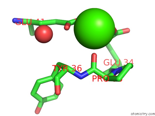
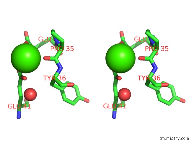
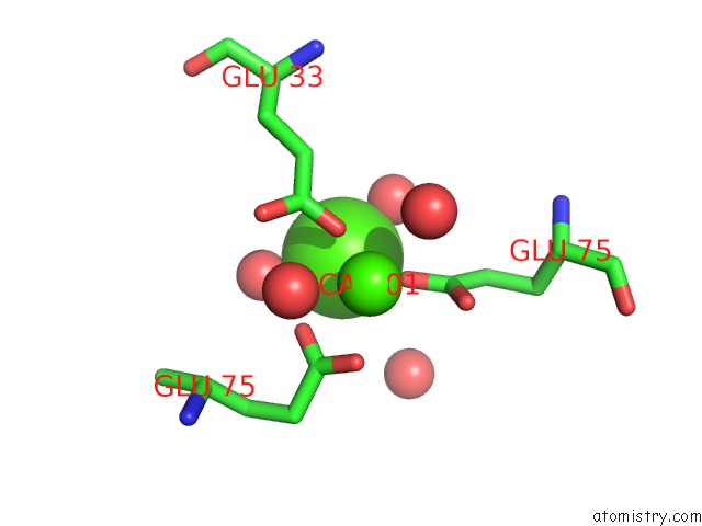
Calcium binding site 2 out of 9 in 5ug4
Go back to
Calcium binding site 2 out
of 9 in the Structure of Spermidine N-Acetyltransferase Speg From Vibrio Cholerae
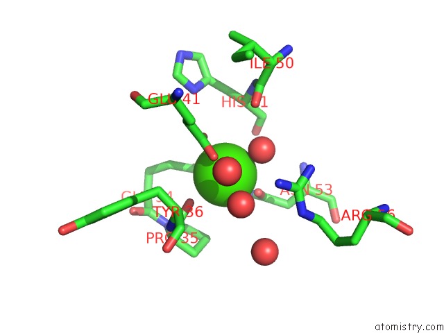
Mono view
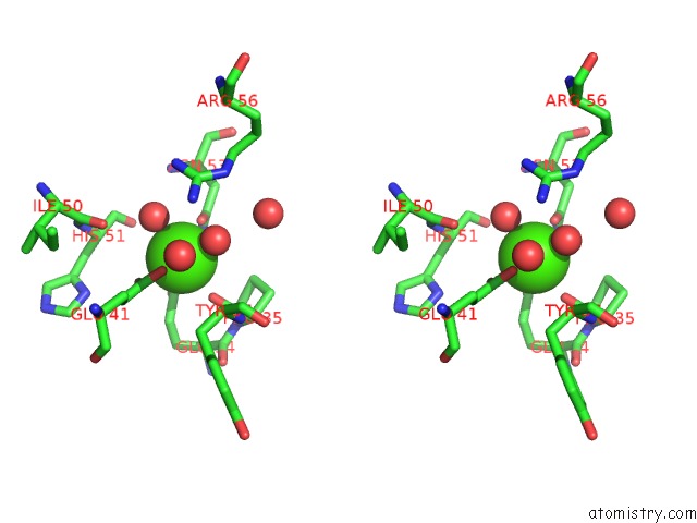
Stereo pair view
Mono view
Stereo pair view
A full contact list of Calcium with other atoms in the Ca binding
site number 2 of Structure of Spermidine N-Acetyltransferase Speg From Vibrio Cholerae within 5.0Å range:
|
Calcium binding site 3 out of 9 in 5ug4
Go back to
Calcium binding site 3 out
of 9 in the Structure of Spermidine N-Acetyltransferase Speg From Vibrio Cholerae

Mono view

Stereo pair view
Mono view
Stereo pair view
A full contact list of Calcium with other atoms in the Ca binding
site number 3 of Structure of Spermidine N-Acetyltransferase Speg From Vibrio Cholerae within 5.0Å range:
|
Calcium binding site 4 out of 9 in 5ug4
Go back to
Calcium binding site 4 out
of 9 in the Structure of Spermidine N-Acetyltransferase Speg From Vibrio Cholerae
Mono view
Stereo pair view
Mono view
Stereo pair view
A full contact list of Calcium with other atoms in the Ca binding
site number 4 of Structure of Spermidine N-Acetyltransferase Speg From Vibrio Cholerae within 5.0Å range:
|
Calcium binding site 5 out of 9 in 5ug4
Go back to
Calcium binding site 5 out
of 9 in the Structure of Spermidine N-Acetyltransferase Speg From Vibrio Cholerae
Mono view

Stereo pair view
Mono view
Stereo pair view
A full contact list of Calcium with other atoms in the Ca binding
site number 5 of Structure of Spermidine N-Acetyltransferase Speg From Vibrio Cholerae within 5.0Å range:
|
Calcium binding site 6 out of 9 in 5ug4
Go back to
Calcium binding site 6 out
of 9 in the Structure of Spermidine N-Acetyltransferase Speg From Vibrio Cholerae
Mono view
Stereo pair view
Mono view
Stereo pair view
A full contact list of Calcium with other atoms in the Ca binding
site number 6 of Structure of Spermidine N-Acetyltransferase Speg From Vibrio Cholerae within 5.0Å range:
|
Calcium binding site 7 out of 9 in 5ug4
Go back to
Calcium binding site 7 out
of 9 in the Structure of Spermidine N-Acetyltransferase Speg From Vibrio Cholerae
Mono view
Stereo pair view
Mono view
Stereo pair view
A full contact list of Calcium with other atoms in the Ca binding
site number 7 of Structure of Spermidine N-Acetyltransferase Speg From Vibrio Cholerae within 5.0Å range:
|
Calcium binding site 8 out of 9 in 5ug4
Go back to
Calcium binding site 8 out
of 9 in the Structure of Spermidine N-Acetyltransferase Speg From Vibrio Cholerae
Mono view

Stereo pair view
Mono view
Stereo pair view
A full contact list of Calcium with other atoms in the Ca binding
site number 8 of Structure of Spermidine N-Acetyltransferase Speg From Vibrio Cholerae within 5.0Å range:
|
Calcium binding site 9 out of 9 in 5ug4
Go back to
Calcium binding site 9 out
of 9 in the Structure of Spermidine N-Acetyltransferase Speg From Vibrio Cholerae

Mono view

Stereo pair view
Mono view
Stereo pair view
A full contact list of Calcium with other atoms in the Ca binding
site number 9 of Structure of Spermidine N-Acetyltransferase Speg From Vibrio Cholerae within 5.0Å range:
|
Reference:
E.V.Filippova,
G.Minasov,
L.Shuvalova,
O.Kiryukhina,
W.F.Anderson,
Center For Structural Genomics Of Infectious Diseases(Csgid).
Structure of Spermidine N-Acetyltransferase Speg From Vibrio Cholerae To Be Published.
Page generated: Wed Jul 9 10:29:58 2025
Last articles
F in 4D6TF in 4D5H
F in 4D42
F in 4D43
F in 4D4V
F in 4D3B
F in 4D3A
F in 4D38
F in 4D37
F in 4D33