Calcium »
PDB 5ufq-5uw6 »
5ulv »
Calcium in PDB 5ulv: Malate Dehydrogenase From Methylobacterium Extorquens
Enzymatic activity of Malate Dehydrogenase From Methylobacterium Extorquens
All present enzymatic activity of Malate Dehydrogenase From Methylobacterium Extorquens:
1.1.1.37;
1.1.1.37;
Protein crystallography data
The structure of Malate Dehydrogenase From Methylobacterium Extorquens, PDB code: 5ulv
was solved by
J.M.Gonzalez,
with X-Ray Crystallography technique. A brief refinement statistics is given in the table below:
| Resolution Low / High (Å) | 37.75 / 1.66 |
| Space group | P 64 2 2 |
| Cell size a, b, c (Å), α, β, γ (°) | 108.995, 108.995, 104.659, 90.00, 90.00, 120.00 |
| R / Rfree (%) | 14 / 16.8 |
Calcium Binding Sites:
The binding sites of Calcium atom in the Malate Dehydrogenase From Methylobacterium Extorquens
(pdb code 5ulv). This binding sites where shown within
5.0 Angstroms radius around Calcium atom.
In total 2 binding sites of Calcium where determined in the Malate Dehydrogenase From Methylobacterium Extorquens, PDB code: 5ulv:
Jump to Calcium binding site number: 1; 2;
In total 2 binding sites of Calcium where determined in the Malate Dehydrogenase From Methylobacterium Extorquens, PDB code: 5ulv:
Jump to Calcium binding site number: 1; 2;
Calcium binding site 1 out of 2 in 5ulv
Go back to
Calcium binding site 1 out
of 2 in the Malate Dehydrogenase From Methylobacterium Extorquens
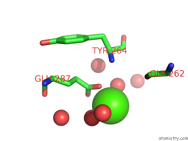
Mono view

Stereo pair view
Mono view
Stereo pair view
A full contact list of Calcium with other atoms in the Ca binding
site number 1 of Malate Dehydrogenase From Methylobacterium Extorquens within 5.0Å range:
|
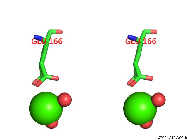
Calcium binding site 2 out of 2 in 5ulv
Go back to
Calcium binding site 2 out
of 2 in the Malate Dehydrogenase From Methylobacterium Extorquens

Mono view
Stereo pair view
Mono view
Stereo pair view
A full contact list of Calcium with other atoms in the Ca binding
site number 2 of Malate Dehydrogenase From Methylobacterium Extorquens within 5.0Å range:
|
Reference:
J.M.Gonzalez,
R.Marti-Arbona,
J.C.H.Chen,
B.Broom-Peltz,
C.J.Unkefer.
Conformational Changes on Substrate Binding Revealed By Structures of Methylobacterium Extorquens Malate Dehydrogenase. Acta Crystallogr F Struct V. 74 610 2018BIOL Commun.
ISSN: ESSN 2053-230X
PubMed: 30279311
DOI: 10.1107/S2053230X18011809
Page generated: Wed Jul 9 10:32:05 2025
ISSN: ESSN 2053-230X
PubMed: 30279311
DOI: 10.1107/S2053230X18011809
Last articles
Cl in 5QE9Cl in 5QCH
Cl in 5QD5
Cl in 5QE6
Cl in 5QCN
Cl in 5QCM
Cl in 5QCG
Cl in 5QCL
Cl in 5QCK
Cl in 5QCE