Calcium »
PDB 5uwk-5vlk »
5vgt »
Calcium in PDB 5vgt: X-Ray Structure of Bacteriophage SF6 Tail Adaptor Protein GP7
Protein crystallography data
The structure of X-Ray Structure of Bacteriophage SF6 Tail Adaptor Protein GP7, PDB code: 5vgt
was solved by
L.Tang,
L.Liang,
H.Zhao,
with X-Ray Crystallography technique. A brief refinement statistics is given in the table below:
| Resolution Low / High (Å) | 19.95 / 1.78 |
| Space group | P 41 21 2 |
| Cell size a, b, c (Å), α, β, γ (°) | 101.297, 101.297, 76.465, 90.00, 90.00, 90.00 |
| R / Rfree (%) | 19.5 / 23.4 |
Other elements in 5vgt:
The structure of X-Ray Structure of Bacteriophage SF6 Tail Adaptor Protein GP7 also contains other interesting chemical elements:
| Magnesium | (Mg) | 1 atom |
Calcium Binding Sites:
The binding sites of Calcium atom in the X-Ray Structure of Bacteriophage SF6 Tail Adaptor Protein GP7
(pdb code 5vgt). This binding sites where shown within
5.0 Angstroms radius around Calcium atom.
In total 4 binding sites of Calcium where determined in the X-Ray Structure of Bacteriophage SF6 Tail Adaptor Protein GP7, PDB code: 5vgt:
Jump to Calcium binding site number: 1; 2; 3; 4;
In total 4 binding sites of Calcium where determined in the X-Ray Structure of Bacteriophage SF6 Tail Adaptor Protein GP7, PDB code: 5vgt:
Jump to Calcium binding site number: 1; 2; 3; 4;
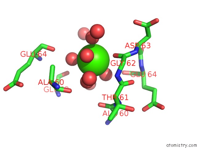
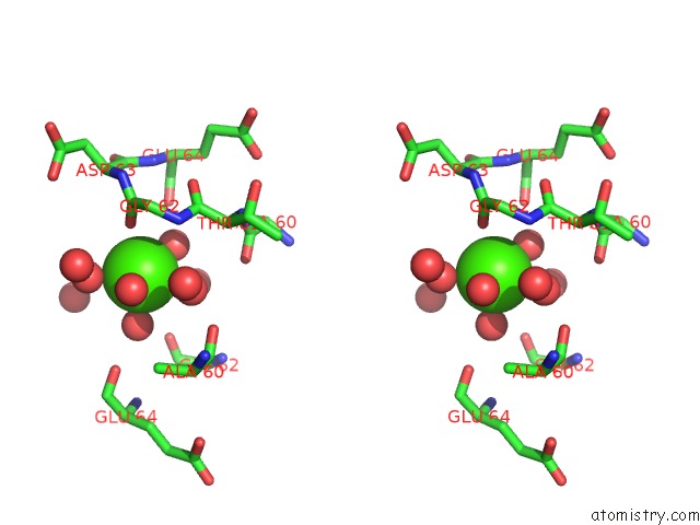
Calcium binding site 1 out of 4 in 5vgt
Go back to
Calcium binding site 1 out
of 4 in the X-Ray Structure of Bacteriophage SF6 Tail Adaptor Protein GP7
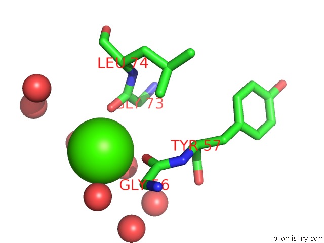
Mono view
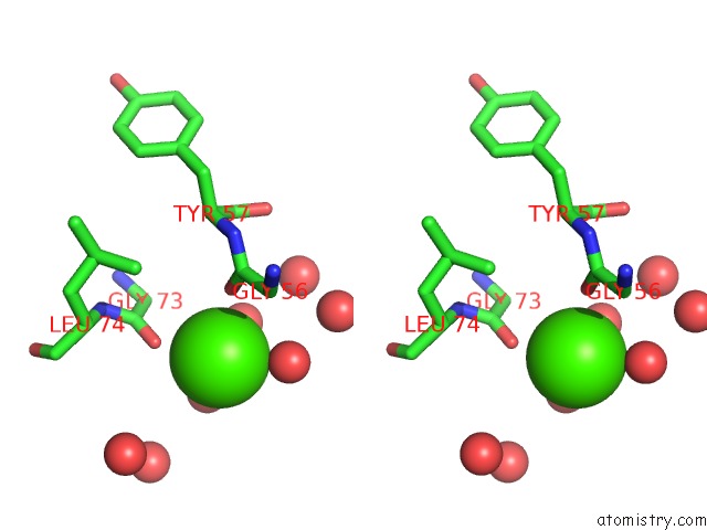
Stereo pair view
Mono view
Stereo pair view
A full contact list of Calcium with other atoms in the Ca binding
site number 1 of X-Ray Structure of Bacteriophage SF6 Tail Adaptor Protein GP7 within 5.0Å range:
|
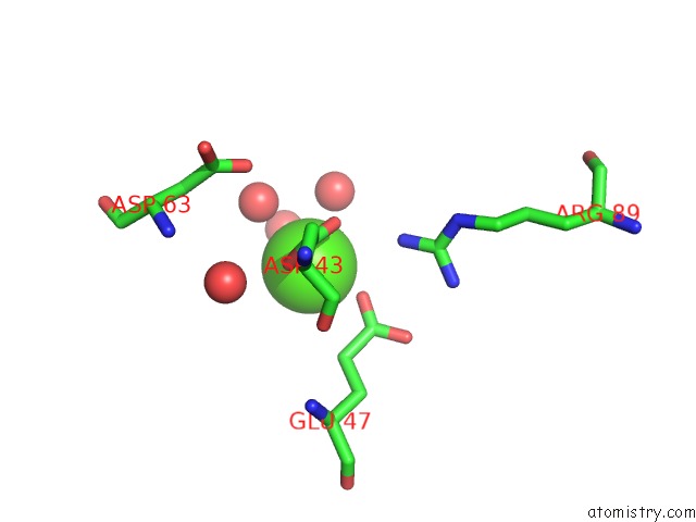
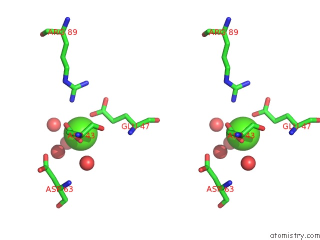
Calcium binding site 2 out of 4 in 5vgt
Go back to
Calcium binding site 2 out
of 4 in the X-Ray Structure of Bacteriophage SF6 Tail Adaptor Protein GP7
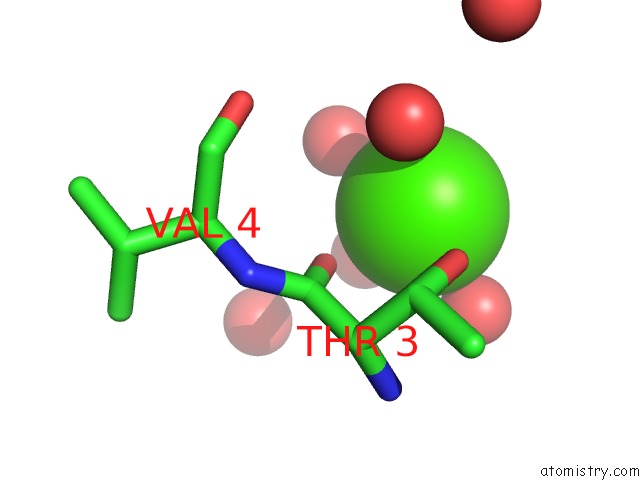
Mono view
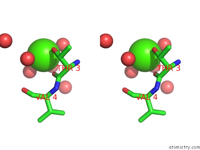
Stereo pair view
Mono view
Stereo pair view
A full contact list of Calcium with other atoms in the Ca binding
site number 2 of X-Ray Structure of Bacteriophage SF6 Tail Adaptor Protein GP7 within 5.0Å range:
|
Calcium binding site 3 out of 4 in 5vgt
Go back to
Calcium binding site 3 out
of 4 in the X-Ray Structure of Bacteriophage SF6 Tail Adaptor Protein GP7
Mono view
Stereo pair view
Mono view
Stereo pair view
A full contact list of Calcium with other atoms in the Ca binding
site number 3 of X-Ray Structure of Bacteriophage SF6 Tail Adaptor Protein GP7 within 5.0Å range:
|
Calcium binding site 4 out of 4 in 5vgt
Go back to
Calcium binding site 4 out
of 4 in the X-Ray Structure of Bacteriophage SF6 Tail Adaptor Protein GP7
Mono view
Stereo pair view
Mono view
Stereo pair view
A full contact list of Calcium with other atoms in the Ca binding
site number 4 of X-Ray Structure of Bacteriophage SF6 Tail Adaptor Protein GP7 within 5.0Å range:
|
Reference:
L.Liang,
H.Zhao,
B.An,
L.Tang.
High-Resolution Structure of Podovirus Tail Adaptor Suggests Repositioning of An Octad Motif That Mediates the Sequential Tail Assembly. Proc. Natl. Acad. Sci. V. 115 313 2018U.S.A..
ISSN: ESSN 1091-6490
PubMed: 29279385
DOI: 10.1073/PNAS.1706846115
Page generated: Mon Jul 15 12:26:16 2024
ISSN: ESSN 1091-6490
PubMed: 29279385
DOI: 10.1073/PNAS.1706846115
Last articles
Zn in 9J0NZn in 9J0O
Zn in 9J0P
Zn in 9FJX
Zn in 9EKB
Zn in 9C0F
Zn in 9CAH
Zn in 9CH0
Zn in 9CH3
Zn in 9CH1