Calcium »
PDB 5vll-5w78 »
5vyb »
Calcium in PDB 5vyb: Structure of the Carbohydrate Recognition Domain of Dectin-2 Complexed with A Mammalian-Type High Mannose MAN9GLCNAC2 Oligosaccharide
Protein crystallography data
The structure of Structure of the Carbohydrate Recognition Domain of Dectin-2 Complexed with A Mammalian-Type High Mannose MAN9GLCNAC2 Oligosaccharide, PDB code: 5vyb
was solved by
H.Feinberg,
S.A.F.Jegouzo,
M.J.Rex,
K.Drickamer,
M.E.Taylor,
W.I.Weis,
with X-Ray Crystallography technique. A brief refinement statistics is given in the table below:
| Resolution Low / High (Å) | 38.23 / 2.40 |
| Space group | C 2 2 21 |
| Cell size a, b, c (Å), α, β, γ (°) | 64.055, 78.834, 76.459, 90.00, 90.00, 90.00 |
| R / Rfree (%) | 20.5 / 24 |
Other elements in 5vyb:
The structure of Structure of the Carbohydrate Recognition Domain of Dectin-2 Complexed with A Mammalian-Type High Mannose MAN9GLCNAC2 Oligosaccharide also contains other interesting chemical elements:
| Sodium | (Na) | 1 atom |
Calcium Binding Sites:
The binding sites of Calcium atom in the Structure of the Carbohydrate Recognition Domain of Dectin-2 Complexed with A Mammalian-Type High Mannose MAN9GLCNAC2 Oligosaccharide
(pdb code 5vyb). This binding sites where shown within
5.0 Angstroms radius around Calcium atom.
In total 2 binding sites of Calcium where determined in the Structure of the Carbohydrate Recognition Domain of Dectin-2 Complexed with A Mammalian-Type High Mannose MAN9GLCNAC2 Oligosaccharide, PDB code: 5vyb:
Jump to Calcium binding site number: 1; 2;
In total 2 binding sites of Calcium where determined in the Structure of the Carbohydrate Recognition Domain of Dectin-2 Complexed with A Mammalian-Type High Mannose MAN9GLCNAC2 Oligosaccharide, PDB code: 5vyb:
Jump to Calcium binding site number: 1; 2;
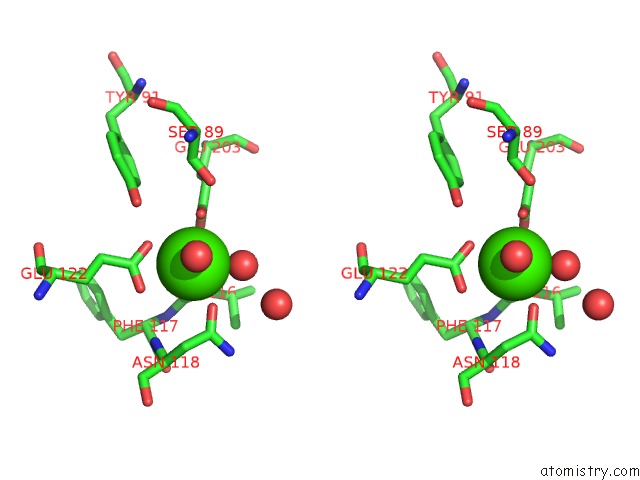
Calcium binding site 1 out of 2 in 5vyb
Go back to
Calcium binding site 1 out
of 2 in the Structure of the Carbohydrate Recognition Domain of Dectin-2 Complexed with A Mammalian-Type High Mannose MAN9GLCNAC2 Oligosaccharide

Mono view

Stereo pair view
Mono view
Stereo pair view
A full contact list of Calcium with other atoms in the Ca binding
site number 1 of Structure of the Carbohydrate Recognition Domain of Dectin-2 Complexed with A Mammalian-Type High Mannose MAN9GLCNAC2 Oligosaccharide within 5.0Å range:
|
Calcium binding site 2 out of 2 in 5vyb
Go back to
Calcium binding site 2 out
of 2 in the Structure of the Carbohydrate Recognition Domain of Dectin-2 Complexed with A Mammalian-Type High Mannose MAN9GLCNAC2 Oligosaccharide

Mono view
Stereo pair view
Mono view
Stereo pair view
A full contact list of Calcium with other atoms in the Ca binding
site number 2 of Structure of the Carbohydrate Recognition Domain of Dectin-2 Complexed with A Mammalian-Type High Mannose MAN9GLCNAC2 Oligosaccharide within 5.0Å range:
|
Reference:
H.Feinberg,
S.A.F.Jegouzo,
M.J.Rex,
K.Drickamer,
W.I.Weis,
M.E.Taylor.
Mechanism of Pathogen Recognition By Human Dectin-2. J. Biol. Chem. V. 292 13402 2017.
ISSN: ESSN 1083-351X
PubMed: 28652405
DOI: 10.1074/JBC.M117.799080
Page generated: Mon Jul 15 12:39:52 2024
ISSN: ESSN 1083-351X
PubMed: 28652405
DOI: 10.1074/JBC.M117.799080
Last articles
Zn in 9MJ5Zn in 9HNW
Zn in 9G0L
Zn in 9FNE
Zn in 9DZN
Zn in 9E0I
Zn in 9D32
Zn in 9DAK
Zn in 8ZXC
Zn in 8ZUF