Calcium »
PDB 5vll-5w78 »
5vyg »
Calcium in PDB 5vyg: Crystal Structure of HFA9 Egf Repeat with O-Glucose Trisaccharide
Enzymatic activity of Crystal Structure of HFA9 Egf Repeat with O-Glucose Trisaccharide
All present enzymatic activity of Crystal Structure of HFA9 Egf Repeat with O-Glucose Trisaccharide:
3.4.21.22;
3.4.21.22;
Protein crystallography data
The structure of Crystal Structure of HFA9 Egf Repeat with O-Glucose Trisaccharide, PDB code: 5vyg
was solved by
H.J.Yu,
H.L.Li,
with X-Ray Crystallography technique. A brief refinement statistics is given in the table below:
| Resolution Low / High (Å) | 46.16 / 2.20 |
| Space group | C 1 2 1 |
| Cell size a, b, c (Å), α, β, γ (°) | 92.417, 46.840, 37.259, 90.00, 92.58, 90.00 |
| R / Rfree (%) | 21.2 / 25.9 |
Calcium Binding Sites:
The binding sites of Calcium atom in the Crystal Structure of HFA9 Egf Repeat with O-Glucose Trisaccharide
(pdb code 5vyg). This binding sites where shown within
5.0 Angstroms radius around Calcium atom.
In total 3 binding sites of Calcium where determined in the Crystal Structure of HFA9 Egf Repeat with O-Glucose Trisaccharide, PDB code: 5vyg:
Jump to Calcium binding site number: 1; 2; 3;
In total 3 binding sites of Calcium where determined in the Crystal Structure of HFA9 Egf Repeat with O-Glucose Trisaccharide, PDB code: 5vyg:
Jump to Calcium binding site number: 1; 2; 3;
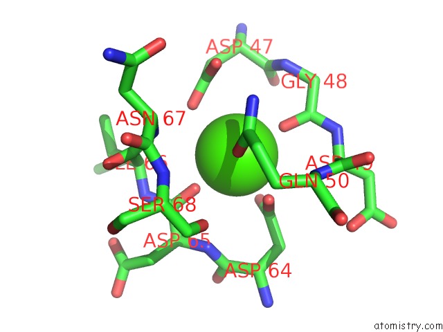
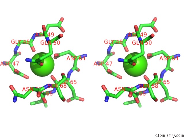
Calcium binding site 1 out of 3 in 5vyg
Go back to
Calcium binding site 1 out
of 3 in the Crystal Structure of HFA9 Egf Repeat with O-Glucose Trisaccharide
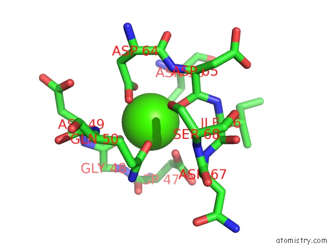
Mono view
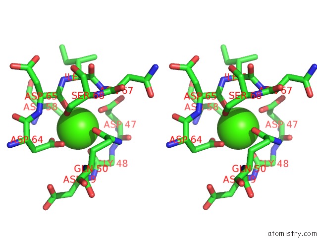
Stereo pair view
Mono view
Stereo pair view
A full contact list of Calcium with other atoms in the Ca binding
site number 1 of Crystal Structure of HFA9 Egf Repeat with O-Glucose Trisaccharide within 5.0Å range:
|
Calcium binding site 2 out of 3 in 5vyg
Go back to
Calcium binding site 2 out
of 3 in the Crystal Structure of HFA9 Egf Repeat with O-Glucose Trisaccharide
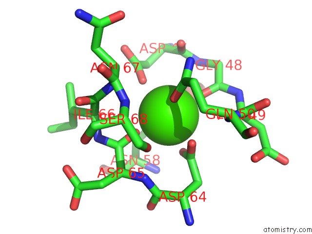
Mono view
Stereo pair view
Mono view
Stereo pair view
A full contact list of Calcium with other atoms in the Ca binding
site number 2 of Crystal Structure of HFA9 Egf Repeat with O-Glucose Trisaccharide within 5.0Å range:
|
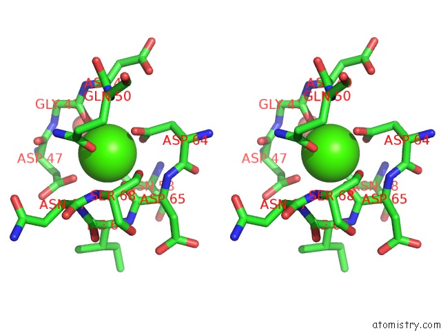
Calcium binding site 3 out of 3 in 5vyg
Go back to
Calcium binding site 3 out
of 3 in the Crystal Structure of HFA9 Egf Repeat with O-Glucose Trisaccharide
Mono view
Stereo pair view
Mono view
Stereo pair view
A full contact list of Calcium with other atoms in the Ca binding
site number 3 of Crystal Structure of HFA9 Egf Repeat with O-Glucose Trisaccharide within 5.0Å range:
|
Reference:
H.Takeuchi,
H.Yu,
H.Hao,
M.Takeuchi,
A.Ito,
H.Li,
R.S.Haltiwanger.
O-Glycosylation Modulates the Stability of Epidermal Growth Factor-Like Repeats and Thereby Regulates Notch Trafficking J. Biol. Chem. V. 292 15964 2017.
ISSN: ESSN 1083-351X
PubMed: 28729422
DOI: 10.1074/JBC.M117.800102
Page generated: Wed Jul 9 10:55:16 2025
ISSN: ESSN 1083-351X
PubMed: 28729422
DOI: 10.1074/JBC.M117.800102
Last articles
Fe in 2YXOFe in 2YRS
Fe in 2YXC
Fe in 2YNM
Fe in 2YVJ
Fe in 2YP1
Fe in 2YU2
Fe in 2YU1
Fe in 2YQB
Fe in 2YOO