Calcium in PDB 5w1d: Crystal Structure of Mouse Protocadherin-15 EC4-7
Protein crystallography data
The structure of Crystal Structure of Mouse Protocadherin-15 EC4-7, PDB code: 5w1d
was solved by
C.F.Klanseck,
B.L.Neel,
M.Sotomayor,
with X-Ray Crystallography technique. A brief refinement statistics is given in the table below:
Resolution Low / High (Å)
|
76.09 /
3.35
|
Space group
|
P 43 21 2
|
Cell size a, b, c (Å), α, β, γ (°)
|
78.863,
78.863,
289.469,
90.00,
90.00,
90.00
|
R / Rfree (%)
|
22.7 /
27.9
|
Other elements in 5w1d:
The structure of Crystal Structure of Mouse Protocadherin-15 EC4-7 also contains other interesting chemical elements:
Calcium Binding Sites:
The binding sites of Calcium atom in the Crystal Structure of Mouse Protocadherin-15 EC4-7
(pdb code 5w1d). This binding sites where shown within
5.0 Angstroms radius around Calcium atom.
In total 6 binding sites of Calcium where determined in the
Crystal Structure of Mouse Protocadherin-15 EC4-7, PDB code: 5w1d:
Jump to Calcium binding site number:
1;
2;
3;
4;
5;
6;
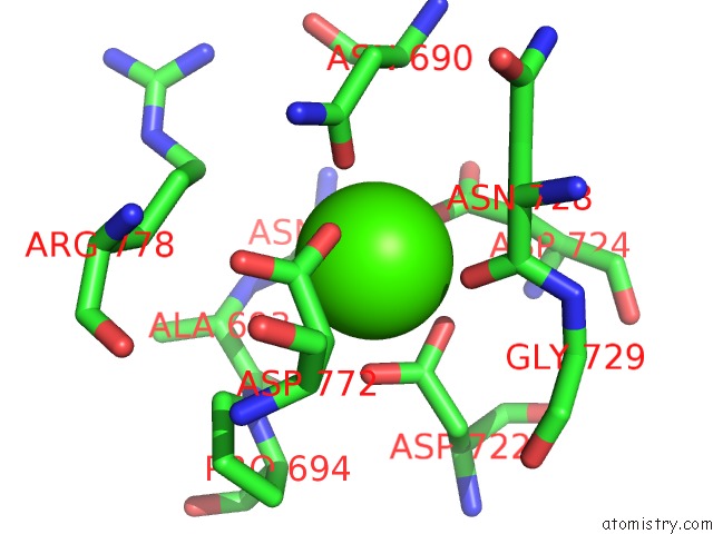
Calcium binding site 1 out
of 6 in 5w1d
Go back to
Calcium Binding Sites List in 5w1d
Calcium binding site 1 out
of 6 in the Crystal Structure of Mouse Protocadherin-15 EC4-7
 Mono view
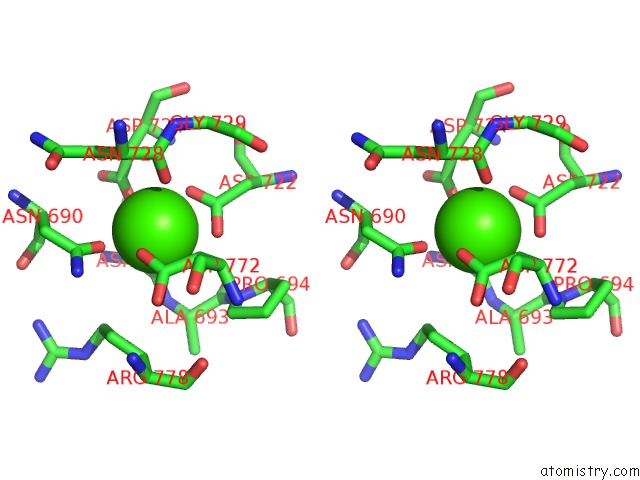
 Stereo pair view
|
A full contact list of Calcium with other atoms in the Ca binding
site number 1 of Crystal Structure of Mouse Protocadherin-15 EC4-7 within 5.0Å range:
probe
|
atom
|
residue
|
distance (Å)
|
B
|
Occ
|
A:Ca1001
b:0.5
occ:1.00
|
OD2
|
A:ASP724
|
2.3
|
95.6
|
1.0
|
O
|
A:ASN692
|
2.3
|
0.1
|
1.0
|
OD1
|
A:ASP722
|
2.4
|
0.9
|
1.0
|
OD1
|
A:ASN690
|
2.4
|
0.8
|
1.0
|
OD2
|
A:ASP722
|
2.6
|
0.3
|
1.0
|
OD2
|
A:ASP772
|
2.6
|
0.6
|
1.0
|
O
|
A:ASN728
|
2.6
|
0.2
|
1.0
|
CG
|
A:ASP722
|
2.8
|
0.7
|
1.0
|
CG
|
A:ASP772
|
3.4
|
0.4
|
1.0
|
CG
|
A:ASP724
|
3.4
|
0.6
|
1.0
|
CG
|
A:ASN690
|
3.5
|
0.9
|
1.0
|
C
|
A:ASN692
|
3.5
|
0.8
|
1.0
|
CB
|
A:ASP772
|
3.6
|
0.8
|
1.0
|
C
|
A:ASN728
|
3.8
|
0.7
|
1.0
|
CB
|
A:ASP724
|
3.9
|
0.8
|
1.0
|
ND2
|
A:ASN690
|
4.1
|
0.1
|
1.0
|
CB
|
A:ASP722
|
4.3
|
0.1
|
1.0
|
CA
|
A:ASN692
|
4.3
|
0.6
|
1.0
|
N
|
A:ASN692
|
4.4
|
0.3
|
1.0
|
CA
|
A:ASN728
|
4.4
|
0.3
|
1.0
|
CB
|
A:ASN728
|
4.4
|
0.9
|
1.0
|
OD1
|
A:ASP772
|
4.4
|
0.3
|
1.0
|
OD1
|
A:ASP724
|
4.4
|
0.7
|
1.0
|
N
|
A:ALA693
|
4.5
|
0.1
|
1.0
|
CB
|
A:ASN690
|
4.6
|
0.1
|
1.0
|
CB
|
A:ASN692
|
4.6
|
0.6
|
1.0
|
CA
|
A:ALA693
|
4.6
|
0.1
|
1.0
|
CD
|
A:PRO694
|
4.7
|
0.5
|
1.0
|
N
|
A:PRO694
|
4.7
|
0.8
|
1.0
|
C
|
A:ALA693
|
4.8
|
0.8
|
1.0
|
CG
|
A:ARG778
|
4.8
|
0.6
|
1.0
|
CA
|
A:ASN690
|
4.8
|
0.5
|
1.0
|
CD
|
A:ARG778
|
4.8
|
0.0
|
1.0
|
N
|
A:GLY729
|
4.8
|
0.4
|
1.0
|
|
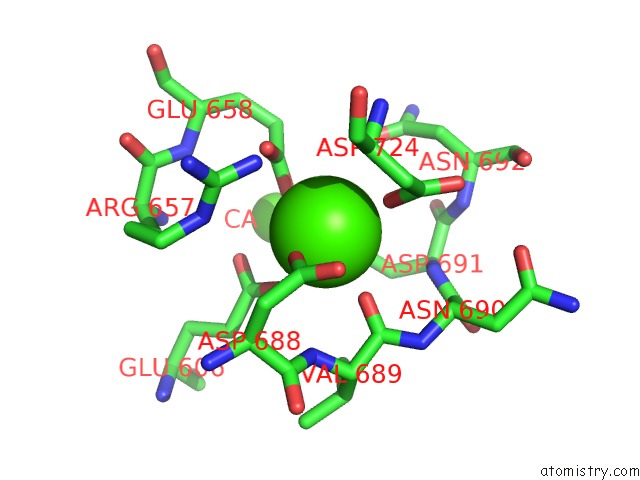
Calcium binding site 2 out
of 6 in 5w1d
Go back to
Calcium Binding Sites List in 5w1d
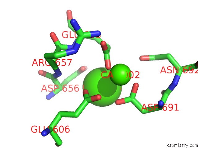
Calcium binding site 2 out
of 6 in the Crystal Structure of Mouse Protocadherin-15 EC4-7
 Mono view
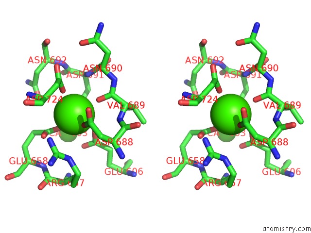
 Stereo pair view
|
A full contact list of Calcium with other atoms in the Ca binding
site number 2 of Crystal Structure of Mouse Protocadherin-15 EC4-7 within 5.0Å range:
probe
|
atom
|
residue
|
distance (Å)
|
B
|
Occ
|
A:Ca1002
b:95.5
occ:1.00
|
OD1
|
A:ASP688
|
2.0
|
97.0
|
1.0
|
OD1
|
A:ASP724
|
2.3
|
0.7
|
1.0
|
OE1
|
A:GLU658
|
2.4
|
0.8
|
1.0
|
O
|
A:VAL689
|
2.5
|
0.7
|
1.0
|
OE2
|
A:GLU606
|
2.6
|
0.0
|
1.0
|
OD1
|
A:ASP691
|
2.7
|
0.2
|
1.0
|
CG
|
A:ASP688
|
3.1
|
0.0
|
1.0
|
CD
|
A:GLU658
|
3.2
|
0.6
|
1.0
|
OE2
|
A:GLU658
|
3.2
|
0.2
|
1.0
|
CG
|
A:ASP691
|
3.4
|
0.2
|
1.0
|
CG
|
A:ASP724
|
3.4
|
0.6
|
1.0
|
OD2
|
A:ASP691
|
3.5
|
0.0
|
1.0
|
OD2
|
A:ASP688
|
3.5
|
0.8
|
1.0
|
CD
|
A:GLU606
|
3.6
|
0.5
|
1.0
|
C
|
A:VAL689
|
3.7
|
1.0
|
1.0
|
CB
|
A:ASP724
|
3.8
|
0.8
|
1.0
|
CA
|
A:ASP724
|
3.9
|
0.4
|
1.0
|
OE1
|
A:GLU606
|
3.9
|
0.2
|
1.0
|
NH2
|
A:ARG657
|
3.9
|
0.8
|
1.0
|
N
|
A:VAL689
|
4.0
|
0.9
|
1.0
|
N
|
A:ASP691
|
4.2
|
0.9
|
1.0
|
NE
|
A:ARG657
|
4.3
|
0.1
|
1.0
|
OD1
|
A:ASN692
|
4.4
|
0.2
|
1.0
|
CB
|
A:ASP688
|
4.4
|
0.4
|
1.0
|
CA
|
A:CA1003
|
4.5
|
0.9
|
1.0
|
OD2
|
A:ASP724
|
4.5
|
95.6
|
1.0
|
CG
|
A:GLU658
|
4.5
|
0.5
|
1.0
|
CA
|
A:VAL689
|
4.6
|
0.9
|
1.0
|
N
|
A:ASP724
|
4.6
|
0.1
|
1.0
|
CB
|
A:ASP691
|
4.6
|
0.5
|
1.0
|
CZ
|
A:ARG657
|
4.6
|
0.8
|
1.0
|
N
|
A:ASN690
|
4.6
|
0.3
|
1.0
|
CA
|
A:ASP688
|
4.7
|
0.7
|
1.0
|
CA
|
A:ASN690
|
4.7
|
0.5
|
1.0
|
CG2
|
A:VAL689
|
4.8
|
0.0
|
1.0
|
C
|
A:ASP688
|
4.9
|
0.1
|
1.0
|
CA
|
A:ASP691
|
4.9
|
0.5
|
1.0
|
C
|
A:ASN690
|
4.9
|
0.4
|
1.0
|
CG
|
A:GLU606
|
5.0
|
0.9
|
1.0
|
C
|
A:ASP724
|
5.0
|
0.9
|
1.0
|
|
Calcium binding site 3 out
of 6 in 5w1d
Go back to
Calcium Binding Sites List in 5w1d
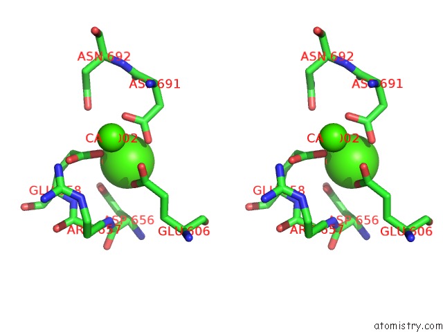
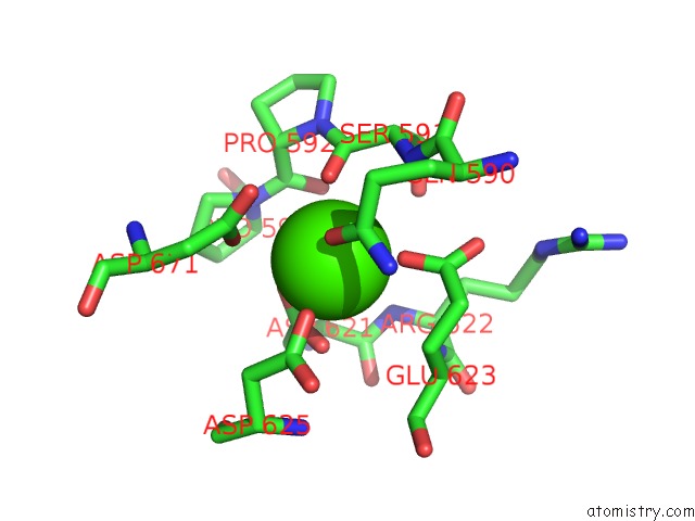
Calcium binding site 3 out
of 6 in the Crystal Structure of Mouse Protocadherin-15 EC4-7
 Mono view
 Stereo pair view
|
A full contact list of Calcium with other atoms in the Ca binding
site number 3 of Crystal Structure of Mouse Protocadherin-15 EC4-7 within 5.0Å range:
probe
|
atom
|
residue
|
distance (Å)
|
B
|
Occ
|
A:Ca1003
b:0.9
occ:1.00
|
OD2
|
A:ASP691
|
2.4
|
0.0
|
1.0
|
OE1
|
A:GLU606
|
2.5
|
0.2
|
1.0
|
OD1
|
A:ASP656
|
2.7
|
0.3
|
1.0
|
OE1
|
A:GLU658
|
2.7
|
0.8
|
1.0
|
CG
|
A:ASP691
|
3.2
|
0.2
|
1.0
|
CG
|
A:ASP656
|
3.4
|
0.2
|
1.0
|
CD
|
A:GLU606
|
3.5
|
0.5
|
1.0
|
OD1
|
A:ASP691
|
3.5
|
0.2
|
1.0
|
CD
|
A:GLU658
|
3.5
|
0.6
|
1.0
|
CG
|
A:GLU658
|
3.6
|
0.5
|
1.0
|
OE2
|
A:GLU606
|
3.9
|
0.0
|
1.0
|
CA
|
A:ASP656
|
4.0
|
0.8
|
1.0
|
OD2
|
A:ASP656
|
4.0
|
0.0
|
1.0
|
CB
|
A:ASP656
|
4.2
|
0.4
|
1.0
|
N
|
A:ARG657
|
4.2
|
0.5
|
1.0
|
CB
|
A:ASP691
|
4.4
|
0.5
|
1.0
|
CA
|
A:CA1002
|
4.5
|
95.5
|
1.0
|
OD1
|
A:ASN692
|
4.5
|
0.2
|
1.0
|
N
|
A:GLU658
|
4.5
|
0.3
|
1.0
|
CB
|
A:GLU658
|
4.6
|
0.2
|
1.0
|
C
|
A:ASP656
|
4.6
|
0.4
|
1.0
|
CG
|
A:GLU606
|
4.6
|
0.9
|
1.0
|
OE2
|
A:GLU658
|
4.7
|
0.2
|
1.0
|
CB
|
A:GLU606
|
5.0
|
0.6
|
1.0
|
|
Calcium binding site 4 out
of 6 in 5w1d
Go back to
Calcium Binding Sites List in 5w1d
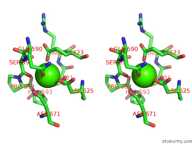
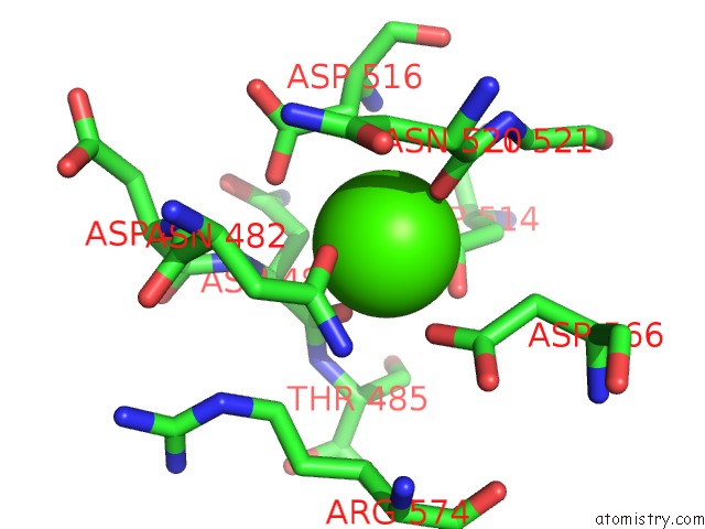
Calcium binding site 4 out
of 6 in the Crystal Structure of Mouse Protocadherin-15 EC4-7
 Mono view
 Stereo pair view
|
A full contact list of Calcium with other atoms in the Ca binding
site number 4 of Crystal Structure of Mouse Protocadherin-15 EC4-7 within 5.0Å range:
probe
|
atom
|
residue
|
distance (Å)
|
B
|
Occ
|
A:Ca1004
b:0.6
occ:1.00
|
OE1
|
A:GLU623
|
2.2
|
0.3
|
1.0
|
OD1
|
A:ASP621
|
2.3
|
0.1
|
1.0
|
OE1
|
A:GLN590
|
2.4
|
0.1
|
1.0
|
O
|
A:SER591
|
2.5
|
0.5
|
1.0
|
OD2
|
A:ASP625
|
2.6
|
0.5
|
1.0
|
OD2
|
A:ASP621
|
2.8
|
0.7
|
1.0
|
CG
|
A:ASP621
|
2.9
|
0.0
|
1.0
|
OD1
|
A:ASP671
|
3.0
|
0.1
|
1.0
|
CD
|
A:GLN590
|
3.4
|
0.9
|
1.0
|
CD
|
A:GLU623
|
3.4
|
0.9
|
1.0
|
CG
|
A:ASP671
|
3.5
|
0.3
|
1.0
|
C
|
A:SER591
|
3.5
|
0.8
|
1.0
|
OD2
|
A:ASP671
|
3.6
|
0.3
|
1.0
|
CG
|
A:ASP625
|
3.7
|
0.1
|
1.0
|
N
|
A:SER591
|
3.8
|
0.7
|
1.0
|
CB
|
A:GLU623
|
4.0
|
0.4
|
1.0
|
NE2
|
A:GLN590
|
4.0
|
0.4
|
1.0
|
OG
|
A:SER591
|
4.0
|
0.5
|
1.0
|
CG
|
A:GLU623
|
4.1
|
0.1
|
1.0
|
O
|
A:PRO592
|
4.1
|
0.2
|
1.0
|
C
|
A:PRO592
|
4.2
|
0.8
|
1.0
|
CA
|
A:SER591
|
4.3
|
0.8
|
1.0
|
CB
|
A:ASP625
|
4.3
|
1.0
|
1.0
|
N
|
A:GLU623
|
4.3
|
0.3
|
1.0
|
CB
|
A:ASP621
|
4.3
|
0.7
|
1.0
|
OE2
|
A:GLU623
|
4.4
|
0.7
|
1.0
|
CG
|
A:GLN590
|
4.4
|
0.3
|
1.0
|
CA
|
A:PRO592
|
4.4
|
0.2
|
1.0
|
N
|
A:PRO592
|
4.4
|
0.8
|
1.0
|
OD1
|
A:ASP625
|
4.6
|
0.6
|
1.0
|
N
|
A:ARG622
|
4.6
|
0.5
|
1.0
|
N
|
A:PRO593
|
4.6
|
0.1
|
1.0
|
CB
|
A:ASP671
|
4.7
|
0.7
|
1.0
|
CA
|
A:GLU623
|
4.7
|
0.6
|
1.0
|
CB
|
A:SER591
|
4.8
|
0.1
|
1.0
|
CA
|
A:ASP621
|
4.8
|
0.4
|
1.0
|
C
|
A:ASP621
|
4.9
|
0.3
|
1.0
|
C
|
A:GLN590
|
4.9
|
0.2
|
1.0
|
|
Calcium binding site 5 out
of 6 in 5w1d
Go back to
Calcium Binding Sites List in 5w1d
Calcium binding site 5 out
of 6 in the Crystal Structure of Mouse Protocadherin-15 EC4-7
 Mono view
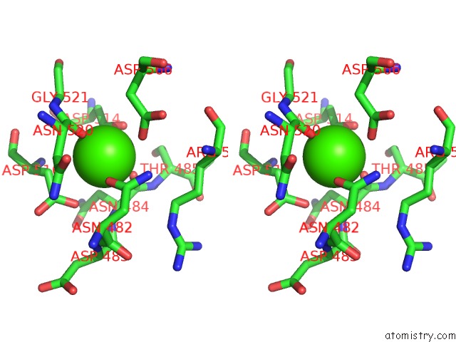
 Stereo pair view
|
A full contact list of Calcium with other atoms in the Ca binding
site number 5 of Crystal Structure of Mouse Protocadherin-15 EC4-7 within 5.0Å range:
probe
|
atom
|
residue
|
distance (Å)
|
B
|
Occ
|
A:Ca1005
b:87.3
occ:1.00
|
O
|
A:ASN520
|
2.1
|
94.0
|
1.0
|
OD1
|
A:ASP514
|
2.2
|
98.5
|
1.0
|
O
|
A:ASN484
|
2.4
|
0.9
|
1.0
|
OD1
|
A:ASN482
|
2.4
|
97.5
|
1.0
|
OD2
|
A:ASP514
|
2.6
|
98.7
|
1.0
|
OD2
|
A:ASP566
|
2.7
|
97.9
|
1.0
|
CG
|
A:ASP514
|
2.7
|
99.0
|
1.0
|
OD2
|
A:ASP516
|
3.2
|
0.7
|
1.0
|
C
|
A:ASN520
|
3.3
|
91.8
|
1.0
|
CB
|
A:ASP516
|
3.4
|
0.1
|
1.0
|
C
|
A:ASN484
|
3.4
|
0.2
|
1.0
|
CG
|
A:ASN482
|
3.5
|
0.8
|
1.0
|
CG
|
A:ASP566
|
3.7
|
0.6
|
1.0
|
CG
|
A:ASP516
|
3.7
|
0.9
|
1.0
|
ND2
|
A:ASN482
|
3.8
|
97.3
|
1.0
|
CB
|
A:ASN484
|
3.9
|
0.7
|
1.0
|
N
|
A:ASN484
|
3.9
|
0.9
|
1.0
|
CA
|
A:ASN484
|
4.0
|
0.6
|
1.0
|
CB
|
A:ASP566
|
4.0
|
0.4
|
1.0
|
CB
|
A:ASN520
|
4.1
|
95.2
|
1.0
|
N
|
A:GLY521
|
4.2
|
89.7
|
1.0
|
CA
|
A:ASN520
|
4.2
|
95.1
|
1.0
|
CB
|
A:ASP514
|
4.2
|
0.1
|
1.0
|
CA
|
A:GLY521
|
4.2
|
90.8
|
1.0
|
N
|
A:THR485
|
4.6
|
0.0
|
1.0
|
CA
|
A:ASP516
|
4.6
|
0.8
|
1.0
|
N
|
A:ASP516
|
4.6
|
0.4
|
1.0
|
OD1
|
A:ASP566
|
4.7
|
0.1
|
1.0
|
CB
|
A:ASN482
|
4.8
|
0.1
|
1.0
|
CD
|
A:ARG574
|
4.9
|
0.3
|
1.0
|
OD1
|
A:ASP516
|
4.9
|
0.2
|
1.0
|
N
|
A:ASP483
|
4.9
|
0.8
|
1.0
|
|
Calcium binding site 6 out
of 6 in 5w1d
Go back to
Calcium Binding Sites List in 5w1d
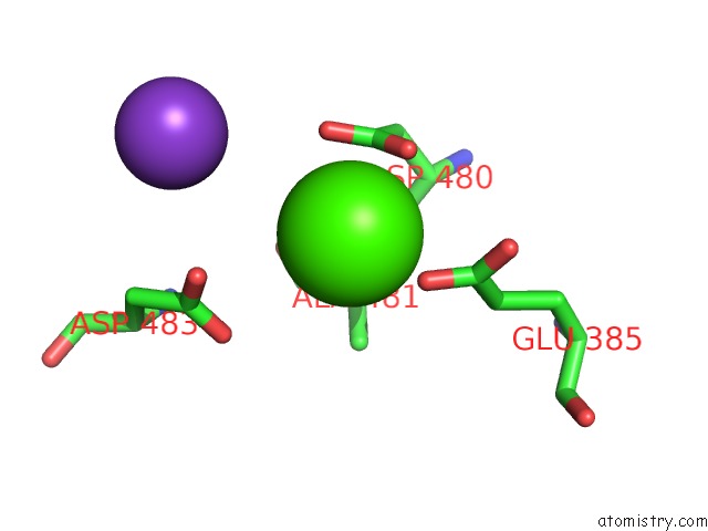
Calcium binding site 6 out
of 6 in the Crystal Structure of Mouse Protocadherin-15 EC4-7
 Mono view
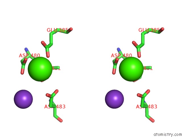
 Stereo pair view
|
A full contact list of Calcium with other atoms in the Ca binding
site number 6 of Crystal Structure of Mouse Protocadherin-15 EC4-7 within 5.0Å range:
probe
|
atom
|
residue
|
distance (Å)
|
B
|
Occ
|
A:Ca1007
b:0.1
occ:1.00
|
OE2
|
A:GLU385
|
2.2
|
0.8
|
1.0
|
OD1
|
A:ASP480
|
2.7
|
0.0
|
1.0
|
CG
|
A:ASP480
|
3.1
|
0.6
|
1.0
|
OD2
|
A:ASP480
|
3.2
|
0.8
|
1.0
|
CD
|
A:GLU385
|
3.3
|
0.8
|
1.0
|
N
|
A:ALA481
|
3.8
|
0.4
|
1.0
|
OE1
|
A:GLU385
|
3.9
|
0.6
|
1.0
|
OD2
|
A:ASP483
|
3.9
|
1.0
|
1.0
|
O
|
A:ALA481
|
4.2
|
0.4
|
1.0
|
CB
|
A:ALA481
|
4.4
|
0.3
|
1.0
|
CB
|
A:ASP480
|
4.4
|
0.4
|
1.0
|
OD1
|
A:ASP483
|
4.4
|
0.8
|
1.0
|
CG
|
A:GLU385
|
4.5
|
0.1
|
1.0
|
CG
|
A:ASP483
|
4.5
|
0.5
|
1.0
|
CA
|
A:ALA481
|
4.5
|
0.0
|
1.0
|
CA
|
A:ASP480
|
4.6
|
0.4
|
1.0
|
C
|
A:ASP480
|
4.6
|
0.4
|
1.0
|
C
|
A:ALA481
|
4.8
|
0.9
|
1.0
|
K
|
A:K1006
|
5.0
|
0.5
|
1.0
|
|
Reference:
C.Klanseck,
B.L.Neel,
M.Sotomayor.
Crystal Structure of Mouse Protocadherin-15 EC4-7 To Be Published.
Page generated: Mon Jul 15 12:41:18 2024
|