Calcium »
PDB 5w7a-5wn3 »
5wd6 »
Calcium in PDB 5wd6: Bovine Salivary Protein Form 30B
Protein crystallography data
The structure of Bovine Salivary Protein Form 30B, PDB code: 5wd6
was solved by
H.Zhang,
V.L.Arcus,
with X-Ray Crystallography technique. A brief refinement statistics is given in the table below:
| Resolution Low / High (Å) | 47.43 / 2.00 |
| Space group | C 1 2 1 |
| Cell size a, b, c (Å), α, β, γ (°) | 81.650, 59.570, 89.940, 90.00, 106.25, 90.00 |
| R / Rfree (%) | 20.5 / 25.1 |
Calcium Binding Sites:
The binding sites of Calcium atom in the Bovine Salivary Protein Form 30B
(pdb code 5wd6). This binding sites where shown within
5.0 Angstroms radius around Calcium atom.
In total 2 binding sites of Calcium where determined in the Bovine Salivary Protein Form 30B, PDB code: 5wd6:
Jump to Calcium binding site number: 1; 2;
In total 2 binding sites of Calcium where determined in the Bovine Salivary Protein Form 30B, PDB code: 5wd6:
Jump to Calcium binding site number: 1; 2;
Calcium binding site 1 out of 2 in 5wd6
Go back to
Calcium binding site 1 out
of 2 in the Bovine Salivary Protein Form 30B
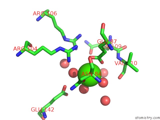
Mono view
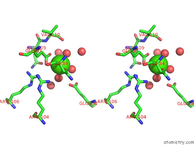
Stereo pair view
Mono view
Stereo pair view
A full contact list of Calcium with other atoms in the Ca binding
site number 1 of Bovine Salivary Protein Form 30B within 5.0Å range:
|
Calcium binding site 2 out of 2 in 5wd6
Go back to
Calcium binding site 2 out
of 2 in the Bovine Salivary Protein Form 30B
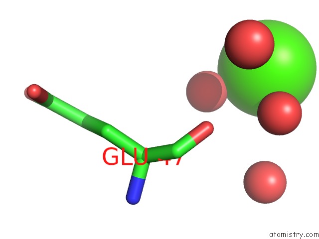
Mono view
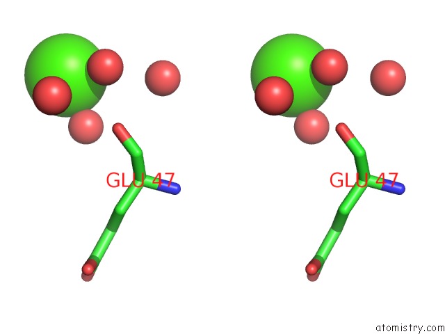
Stereo pair view
Mono view
Stereo pair view
A full contact list of Calcium with other atoms in the Ca binding
site number 2 of Bovine Salivary Protein Form 30B within 5.0Å range:
|
Reference:
H.Zhang,
J.Burrows,
G.L.Card,
G.Attwood,
T.T.Wheeler,
V.L.Arcus.
The Three Dimensional Structure of Bovine Salivary Protein 30B (BSP30B) and Its Interaction with Specific Rumen Bacteria. Plos One V. 14 06709 2019.
ISSN: ESSN 1932-6203
PubMed: 30978191
DOI: 10.1371/JOURNAL.PONE.0206709
Page generated: Mon Jul 15 13:07:18 2024
ISSN: ESSN 1932-6203
PubMed: 30978191
DOI: 10.1371/JOURNAL.PONE.0206709
Last articles
Zn in 9J0NZn in 9J0O
Zn in 9J0P
Zn in 9FJX
Zn in 9EKB
Zn in 9C0F
Zn in 9CAH
Zn in 9CH0
Zn in 9CH3
Zn in 9CH1