Calcium »
PDB 5wzw-5xke »
5x6i »
Calcium in PDB 5x6i: Crystal Structure of B. Subtilis Adenylate Kinase Variant
Enzymatic activity of Crystal Structure of B. Subtilis Adenylate Kinase Variant
All present enzymatic activity of Crystal Structure of B. Subtilis Adenylate Kinase Variant:
2.7.4.3;
2.7.4.3;
Protein crystallography data
The structure of Crystal Structure of B. Subtilis Adenylate Kinase Variant, PDB code: 5x6i
was solved by
S.Moon,
E.Bae,
with X-Ray Crystallography technique. A brief refinement statistics is given in the table below:
| Resolution Low / High (Å) | 50.00 / 2.00 |
| Space group | P 21 21 21 |
| Cell size a, b, c (Å), α, β, γ (°) | 43.739, 44.180, 100.595, 90.00, 90.00, 90.00 |
| R / Rfree (%) | 17.9 / 25.9 |
Other elements in 5x6i:
The structure of Crystal Structure of B. Subtilis Adenylate Kinase Variant also contains other interesting chemical elements:
| Magnesium | (Mg) | 1 atom |
| Zinc | (Zn) | 1 atom |
Calcium Binding Sites:
The binding sites of Calcium atom in the Crystal Structure of B. Subtilis Adenylate Kinase Variant
(pdb code 5x6i). This binding sites where shown within
5.0 Angstroms radius around Calcium atom.
In total 2 binding sites of Calcium where determined in the Crystal Structure of B. Subtilis Adenylate Kinase Variant, PDB code: 5x6i:
Jump to Calcium binding site number: 1; 2;
In total 2 binding sites of Calcium where determined in the Crystal Structure of B. Subtilis Adenylate Kinase Variant, PDB code: 5x6i:
Jump to Calcium binding site number: 1; 2;
Calcium binding site 1 out of 2 in 5x6i
Go back to
Calcium binding site 1 out
of 2 in the Crystal Structure of B. Subtilis Adenylate Kinase Variant
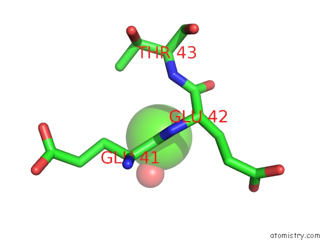
Mono view
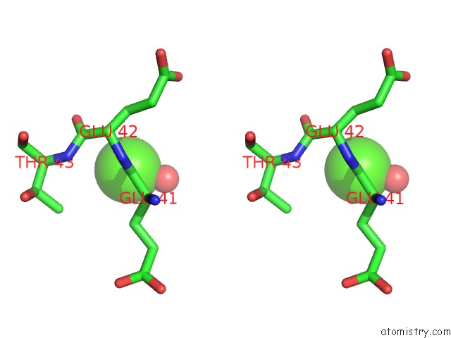
Stereo pair view
Mono view
Stereo pair view
A full contact list of Calcium with other atoms in the Ca binding
site number 1 of Crystal Structure of B. Subtilis Adenylate Kinase Variant within 5.0Å range:
|
Calcium binding site 2 out of 2 in 5x6i
Go back to
Calcium binding site 2 out
of 2 in the Crystal Structure of B. Subtilis Adenylate Kinase Variant
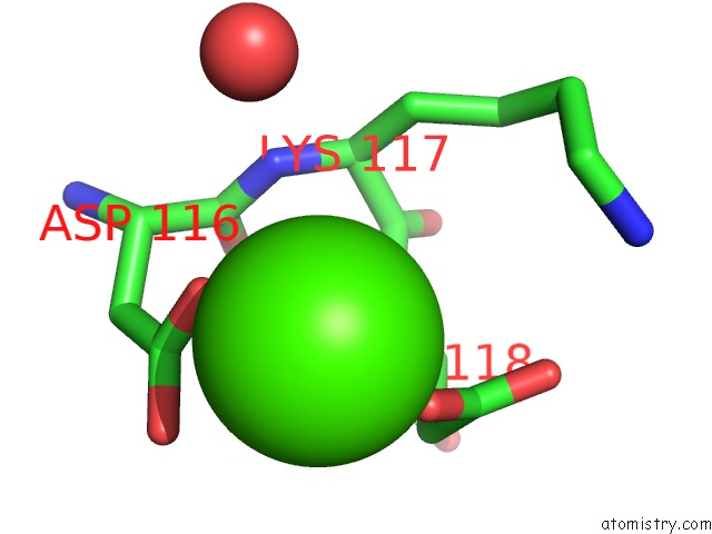
Mono view
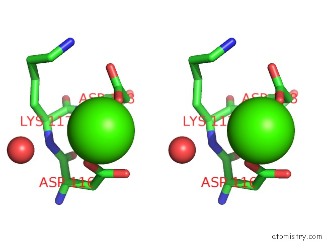
Stereo pair view
Mono view
Stereo pair view
A full contact list of Calcium with other atoms in the Ca binding
site number 2 of Crystal Structure of B. Subtilis Adenylate Kinase Variant within 5.0Å range:
|
Reference:
S.Moon,
J.Kim,
J.Koo,
E.Bae.
Structural and Mutational Analyses of Psychrophilic and Mesophilic Adenylate Kinases Highlight the Role of Hydrophobic Interactions in Protein Thermal Stability. Struct Dyn. V. 6 24702 2019.
ISSN: ESSN 2329-7778
PubMed: 31111079
DOI: 10.1063/1.5089707
Page generated: Wed Jul 9 11:32:02 2025
ISSN: ESSN 2329-7778
PubMed: 31111079
DOI: 10.1063/1.5089707
Last articles
Cl in 5KRTCl in 5KSG
Cl in 5KSF
Cl in 5KS2
Cl in 5KRV
Cl in 5KRL
Cl in 5KRR
Cl in 5KR7
Cl in 5KQK
Cl in 5KQT